Functions of the Nervous and Endocrine Systems
Introduction
The nervous and the endocrine system are both regulatory, but the endocrine system works by using chemical signals whereas the nervous system works by using electrical signal. In this assignment, the anatomy and function of different parts of the nervous and endocrine system is to be discussed.
AC11.1
The structural anatomy of the brain indicates that it is composed of three main parts that are cerebrum, cerebellum and brainstem. The cerebrum is the largest part of the brain and is divided into right and left hemispheres. It supports the functioning of the brain such as interpretation of touch, hearing, vision, formation of speech, reasoning, learning, emotions and fine control of mobilization in individuals (Altamura et al., 2018). The cerebrum is located below the cerebrum and supports the functioning of coordination of muscle movement, maintaining balance and posture. For those pursuing biomedical science, obtaining biomedical science dissertation help can be invaluable in navigating the complexities of these brain functions. The brainstem is the relay centre connecting cerebrum and cerebellum to the spinal cord and it support functions of brain like maintaining heart rate, breathing, wake and sleep cycle, body temperature, coughing, sneezing, digestion, swallowing and vomiting (Herbet and Duffau, 2020).

The spinal cord is cylindrical structure of the body which is made up of white and grey matter. It is uniformly organised and divided into cervical, thoracic, lumbar and sacral regions that are further composed of different segments. It is made up of 31 pairs of spinal nerves out of which 8 cervical nerves, 12 thoracic nerves, 5 lumbar nerves, 5 sacral nerves and 1 coccygeal nerve (Baker and Perez, 2017). The key function of the spinal cord is directing the voluntary muscle movement for moving, allowing electrochemical communication to be established between brain and different parts of the body, maintaining involuntary reflexes and others (Athanasiou et al., 2017).

The peripheral nervous system (PNS) is made up of three parts that are spinal roots, sensory along with autonomic ganglia and the somatic nerves. The peripheral nerves mainly branch out from the brain and spinal cord with the intention of creating communication between the central nervous system and the other parts of the body (Bedbrook et al., 2018). The three key function of PNS is transmitting motor signals to the voluntary striated muscles, carrying external impulse signals from different parts of the brain to the central nervous system and regulation of autonomic functioning like sweating and blood pressure (Jha and Morrison, 2018).

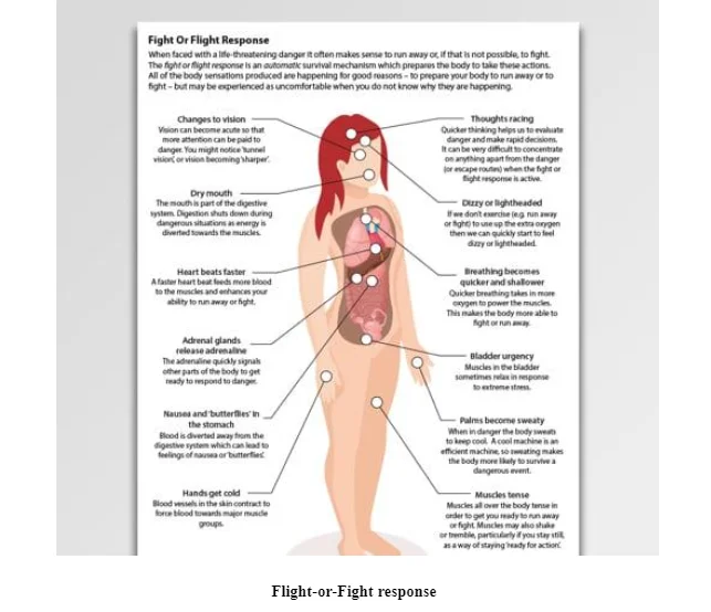
The autonomic nervous system (ANS) is structurally divided into two parts that are sympathetic nervous system and parasympathetic nervous system. The sympathetic division helps in creating flight-or-fight response whereas the parasympathetic division is responsible for ensuring resting and digestion in the body (Sheng and Zhu, 2018). The function of ANS includes regulating rate of breathing, blood pressure, respiration, digestion and others (Sheng and Zhu, 2018).

AC11.2
The reflex arc is referred to the neural pathway which controls the reflexes in the body. The sensory neurons in the vertebrates do not pass through the brain direct but travels through the spinal cord which allows faster reflex action to be performed by activating spinal motor neurons (Costa et al., 2020).
Continue your journey with our comprehensive guide to Overview of the Reproductive System.
The five components of the reflex arc are:
Sensory neuron: The role of the sensory neurons is to transmit the electrical impulse received from the different parts of the body to relay neuron that is located in the spinal cord region. These neurons are mainly activated by the sensory input from the surrounding environment (de Castro et al., 2019).
Sensory receptor: The role of the sensory receptor is responding to the signals or impulse from the environment. The cell on being compressed due to a pressure leads the sensory receptor in the skin to be activated leading the brain to understand to be prepared to respond to a stimulus (de Castro et al., 2019).
Integration centre: The integration centre is composed of multiple synapses including more than one interneuron between motor and sensory neuron. The role of the centre is to act as connecting link for allowing the sensory input to be travelled to other cells of the body in performing action (de Castro et al., 2019).
Motor neuron: The role of the motor neuron is receive signals from the sensory neuron and carry it to the effector muscles to produce response to the stimulus received (de Castro et al., 2019).
Effector target: The effector target has the role to show response received from the motor neurons such as contracting or lifting leg or hands in response to hot feeling (de Castro et al., 2019).

AC11.3
The synaptic transmission is relay of signals from one neuron to another through the axon of the neurons and the propagation of nerve impulse is referred to as jumping of the signals from one node of Ranvier to another (Lee et al., 2018). In response to a nerve impulse, an electrical charge travels along the membrane of a neuron which is generated by the changed membrane potential of the neuron due to exchange of chemical signals. The altered membrane potential leads the nerve cell to be depolarised which is required to reach min of 15 mV to disrupt the resting potential. The depolarisation leads the sodium channels to be opened allowing more sodium ions to enter the cells resulting to increase the positive charge within the cell (+40 mV) initiating action potential. The potassium channel is then seen to open and allow potassium ion to be flown out of the cell to reach the resting potential of -70 mV. The action potential is seen to move in the form of the wave allowing signals to be passed from one node to another node of Ranvier where they are exposed of the myelin sheath (Murphy‐Royal et al., 2017).
The action potential travelling through the axon reaches the pre-synaptic terminal of the neuron. On reaching the end, the voltage-dependent calcium channels are activated leading calcium ions to enter the cell of the synaptic end and increase the extracellular charge to be positive. The binding of the vesicles with the presynaptic membrane leads to release of neurotransmitters in the synaptic cleft which then binds with the post-synaptic receptors leading to activation of sodium ion channels in the post-synaptic end. The action potential as a result of exchange of the ions transmits through the synaptic cleft to the other neuron (Mannan et al., 2019).

AC21.1
The anterior pituitary gland produces major hormones such as growth hormone (hGH), prolactin, thyroid-stimulating hormone (TSH), adrenocorticotropic hormone (ADH), follicle-stimulating hormone (FSH) and luteinizing hormone (LH).The hGH produced lead directly promote the growth and development of the body by stimulating the metabolism of lipids, carbohydrates and protein along with breakdown of triglycerides which are supressed to be taken and be accumulated in circulating lipids (Diana et al., 2020). The indirect action of the hormone on binding with the target cells is activating insulin-like growth factor-1 (IGF-1) which promotes somatic growth and metabolism of nutrients in the body for development (Fisher et al., 2020). The FSH stimulates production of gamete by acting on the gonadal target cell. The transduction of the signals induced by hormone is mediated by the FSH-specific G protein-coupled receptor (FSHR) (Casarini and Crépieux, 2019).
The anterior pituitary releases oxytocin and anti-diuretic hormone (ADH). The oxytocin promotes uterine contraction during childbirth by increasing the concentration of calcium ions in the muscle cells of the uterus responsible for contracting the uterus which leads towards its relaxation (Uvnäs-Moberg et al., 2019). The ADH function to maintain blood pressure, body fluid and blood volume through promotion of water reabsorption in the kidneys. The ADH executes the function by binding with the target receptor cells in the collecting ducts of the kidney where it makes the cells to be permeable for allowing water to be transported back into the circulation through reabsorption (Selvaraja et al., 2020). The thyroid gland produces thyroxine and parathyroid gland produces the parathyroid hormone. The hormones perform their function by creating a docking effect on binding with the protein receptors present in the thyroid-sensitive tissues (Ząbczyńska et al., 2018).

The activities of the endocrine glands are regulated by the interplay of the hormone-releasing and inhibition signals between hypothalamus and pituitary. The interplay is known as the hypothalamic-pituitary axis where the hypothalamus secretes many hormones which controlling the action of pituitary gland. The pituitary gland which is considered as master gland of the body controls the function of many endocrine glands. The pituitary controls the rate of activation of endocrine gland through a feedback lip in which the level of hormones in the blood sends positive or negative signals for slowing or increasing release of hormone from the glands (Oyola and Handa, 2017). For example, the pituitary gland on detecting low level of thyroid hormone in the blood leads the endocrine glands to be instigated through impulse in releasing thyroid-stimulating hormone and vice versa. Thus, through back and forth feedback, the hormone level and activity of the endocrine gland is controlled (Gavrila and Hollenberg, 2019).
The other action which manages the activity of the endocrine gland is neural stimuli causes by external environment of the body (Walling and Rosol, 2019). For example, due to neural stimuli of increased exercise, hormones epinephrine and nor-epinephrine is required to be released from the endocrine gland activity. The neuronal signal for the activation is sent through the sympathetic nervous system which directly stimulates the adrenal medulla to actively produce the stress hormones (Walling and Rosol, 2019). The humoral stimuli also control the activities of endocrine gland. This is evident as lower concentration of insulin in the blood leads the pancreatic gland to be activated in releasing increased amount of insulin in the body (Walling and Rosol, 2019).
AC31.1
During the change in body temperature, the central nervous system (CNS) sends signals to the hypothalamus which sends signals in response to different organs in the body to act accordingly in stabilising body temperature. For example, when the body temperature is increased, sweat glands are activated by the signals from the CNS to release increase sweat to cool down the body. The hypothalamus promotes vasoconstriction and vasodilation for lowering and increase body temperate to normal during the rise and fall of body temperature (Nakamura et al., 2018). The other mechanism involves hormonal thermogenesis in which the thyroid gland promotes release of hormone to increase metabolism in the body that enhances increased amount of heat to be produced to normalise fall in body temperature (Johnson and Kellogg Jr, 2018).
The flight-or-fight response is controlled by the sympathetic nervous system in which the sympathetic nerves stimulates the release of catecholamines from the adrenal glands in the form of adrenalin and nor-adrenalin. They help in creating a burst of energy required for responding to eminent dangers perceived by the body. This is done by increasing the heart rate, blood pressure through increased pumping of blood through the heart, increasing breakdown of fat and enhancing increased use of blood sugar through action of insulin in creating the required energy (Walters, 2020). In blood glucose regulation, the autonomic nervous system mainly acts in stimulating the pancreatic gland to release increase amount of insulin and glucagon in the body. The sympathetic nervous system is promoted through exercise to stimulate production of glucagon and maintain normal blood glucose level. The insulin mainly acts to lower blood glucose level and glucagon acts to increase blood sugar level (Güemes and Georgiou, 2018).
Conclusion
The above discussion mentions that brain and spinal cord forms the central nervous system and they function to provide balance and posture to the body, promote digitation, mobilisation, sensation, vision, speech formation and others. The peripheral and autonomic nervous system are parts of the nervous framework in the body and they function to carry sensory information from brain to other parts of the body and vice-versa. The spinal reflex arc mainly supports the transmission of signal from the spinal cord to the parts of the body. The synaptic transmission and nerve impulse propagation is controlled through action potential and sodium and potassium channel along with calcium channel. The endocrine glands support the action of the hormone by reaching the target sell through the blood stream and the peripheral along with the autonomic nervous system manages the endocrine gland in controlling blood sugar level, body temperature and flight-or-fight response.
Recommendations
The recommendation is that both nervous and endocrine system in the body are to be adequately controlled through use of clinical intervention and treatment so that they can work together in supporting physiological needs functioning of the body. It is also recommended that no harm to the central nervous system, peripheral an autonomic nervous system is to be ensure as otherwise it would disrupt the homeostasis state in the body.
Take a deeper dive into Falls in Dementia Patients with our additional resources.
References
Altamura, A.C., Maggioni, E., Dhanoa, T., Ciappolino, V., Paoli, R.A., Cremaschi, L., Prunas, C., Orsenigo, G., Caletti, E., Cinnante, C.M. and Triulzi, F.M., 2018. The impact of psychosis on brain anatomy in bipolar disorder: a structural MRI study. Journal of affective disorders, 233, pp.100-109.
Athanasiou, A., Klados, M.A., Pandria, N., Foroglou, N., Kavazidi, K.R., Polyzoidis, K. and Bamidis, P.D., 2017. A systematic review of investigations into functional brain connectivity following spinal cord injury. Frontiers in Human Neuroscience, 11, p.517.
Baker, S.N. and Perez, M.A., 2017. Reticulospinal contributions to gross hand function after human spinal cord injury. Journal of Neuroscience, 37(40), pp.9778-9784.
Bedbrook, C.N., Deverman, B.E. and Gradinaru, V., 2018. Viral strategies for targeting the central and peripheral nervous systems. Annual review of neuroscience, 41, pp.323-348.
Casarini, L. and Crépieux, P., 2019. Molecular mechanisms of action of FSH. Frontiers in endocrinology, 10, p.305.
Costa, A.F.B.A., da Veiga Argus, A.P., Pisetta, F.P. and Evangelista, A.G., 2020. Basic background in reflex physiology. Journal of Molecular Pathophysiology, 9(1), pp.1-8.
de Castro, M.V., da Silva, M.V.R., Chiarotto, G.B., Volpe, B.B., Santana, M.H., Luzo, .C.M. and de Oliveira, A.L.R., 2019. Reflex arc recovery after spinal cord dorsal root repair with platelet rich plasma (PRP). Brain research bulletin, 152, pp.212-224.
Diana, J.N., Tao, Y., Du, Q., Wang, M., Kumar, C.U., Wu, F. and Jin, T., 2020. PLGA microspheres of hGH of preserved native state prepared using a self-regulated process. Pharmaceutics, 12(7), p.683.
Fisher, D.M., Pastrak, A., Choe, J., Wajnrajch, M.P. and Cara, J., 2020. OR10-04 Interpretation of Insulin-like Growth Factor-1 (IGF-1) Levels Following Administration of Somatrogon (a Long-acting Human Growth Hormone-hGH-CTP). Journal of the Endocrine Society, 4(Supplement_1), pp.OR10-04.
Gavrila, A. and Hollenberg, A.N., 2019. The hypothalamic-pituitary-thyroid axis: physiological regulation and clinical implications. In The Thyroid and Its Diseases (pp. 13-23). Springer, Cham.
Güemes, A. and Georgiou, P., 2018. Review of the role of the nervous system in glucose homoeostasis and future perspectives towards the management of diabetes. Bioelectronic medicine, 4(1), pp.1-18.
Herbet, G. and Duffau, H., 2020. Revisiting the functional anatomy of the human brain: toward a meta-networking theory of cerebral functions. Physiological reviews, 100(3), pp.1181-1228.
Jha, M.K. and Morrison, B.M., 2018. Glia-neuron energy metabolism in health and diseases: New insights into the role of nervous system metabolic transporters. Experimental neurology, 309, pp.23-31.
Johnson, J.M. and Kellogg Jr, D.L., 2018. Skin vasoconstriction as a heat conservation thermoeffector. Handbook of clinical neurology, 156, pp.175-192.
Lee, A., Hirabayashi, Y., Kwon, S.K., Lewis Jr, T.L. and Polleux, F., 2018. Emerging roles of mitochondria in synaptic transmission and neurodegeneration. Current opinion in physiology, 3, pp.82-93.
Mannan, Z.I., Adhikari, S.P., Yang, C., Budhathoki, R.K., Kim, H. and Chua, L., 2019. Memristive imitation of synaptic transmission and plasticity. IEEE transactions on neural networks and learning systems, 30(11), pp.3458-3470.
Murphy‐Royal, C., Dupuis, J., Groc, L. and Oliet, S.H., 2017. Astroglial glutamate transporters in the brain: regulating neurotransmitter homeostasis and synaptic transmission. Journal of neuroscience research, 95(11), pp.2140-2151.
Nakamura, M., Shintani-Ishida, K. and Ikegaya, H., 2018. 5-HT2A Receptor agonist-induced hyperthermia is induced via vasoconstriction by peripheral 5-HT2A receptors and brown adipose tissue thermogenesis by peripheral serotonin loss at a high ambient temperature. Journal of Pharmacology and Experimental Therapeutics, 367(2), pp.356-362.
Oyola, M.G. and Handa, R.J., 2017. Hypothalamic–pituitary–adrenal and hypothalamic–pituitary–gonadal axes: sex differences in regulation of stress responsivity. Stress, 20(5), pp.476-494.
Selvarajan, R.S., Rahim, R.A., Majlis, B.Y., Gopinath, S.C. and Hamzah, A.A., 2020. Ultrasensitive and Highly Selective Graphene-Based Field-Effect Transistor Biosensor for Anti-Diuretic Hormone Detection. Sensors, 20(9), p.2642.
Sheng, Y. and Zhu, L., 2018. The crosstalk between autonomic nervous system and blood vessels. International journal of physiology, pathophysiology and pharmacology, 10(1), p.17.
Uvnäs-Moberg, K., Ekström-Bergström, A., Berg, M., Buckley, S., Pajalic, Z., Hadjigeorgiou, E., Kotłowska, A., Lengler, L., Kielbratowska, B., Leon-Larios, F. and Magistretti, C.M., 2019. Maternal plasma levels of oxytocin during physiological childbirth–a systematic review with implications for uterine contractions and central actions of oxytocin. BMC pregnancy and childbirth, 19(1), pp.1-17.
Walling, B.E. and Rosol, T.J., 2019. Pathology of the Endocrine System. In Toxicologic Pathology for Non-Pathologists (pp. 537-569). Humana, New York, NY.
Walters, S., 2020. 12.3 Understanding Stress. Psychology-1st Canadian Edition.
Ząbczyńska, M., Kozłowska, K. and Pocheć, E., 2018. Glycosylation in the thyroid gland: vital aspects of glycoprotein function in thyrocyte physiology and thyroid disorders. International journal of molecular sciences, 19(9), p.2792.
Yan, Z. and Rein, B., 2021. Mechanisms of synaptic transmission dysregulation in the prefrontal cortex: pathophysiological implications. Molecular Psychiatry, pp.1-21.
Bibliography
Carvalho, D.P. and Dupuy, C., 2017. Thyroid hormone biosynthesis and release. Molecular and cellular endocrinology, 458, pp.6-15.
Marty, M.S., Borgert, C., Coady, K., Green, R., Levine, S.L., Mihaich, E., Ortego, L., Wheeler, J.R., Yi, K.D. and Zorrilla, L.M., 2018. Distinguishing between endocrine disruption and non-specific effects on endocrine systems. Regulatory Toxicology and Pharmacology, 99, pp.142-158.
Ogawa, S., Liu, X., Shepherd, B.S. and Parhar, I.S., 2018. Ghrelin stimulates growth hormone release from the pituitary via hypothalamic growth hormone-releasing hormone neurons in the cichlid, Oreochromis niloticus. Cell and tissue research, 374(2), pp.349-365.


- 24/7 Customer Support
- 100% Customer Satisfaction
- No Privacy Violation
- Quick Services
- Subject Experts