Cell biology
Section 1:
Discuss the selected characteristics of living cells.
- Cell is basic unit of life that is called biological unit. Unicellular organisation is composed of single cell and the multicellular organism consist of two to many cells (Boiteux et al. 2017).
- A living cell has four important components such as plasma membrane, DNA, ribosome and cytoplasm.
- Plasa membrane is the outer membrane of a living cell that separates it from the outer environment.
- Cytoplasm is referred as the jelly like substance and cellular structures that are suspended into it. The jelly like substance of cytoplasm is referred as cytosol. Cytoplasm is considered as the region except the nucleus inside the plasma membrane (Rout and Field, 2017). Cytoplasm plays important roles in providing structural and functional stability to cells.
- DNA (deoxyribose nucleic acid) is the molecule situated inside the living prokaryotic cell which is involved in carrying genetic information from one generation to the next.
- A living prokaryotic cell also consists of ribosome that is crucial in protein formation inside the cells.
- Growth and development are the most important characteristics of cells. Growth of a living cell depends on its growth factors. Protein in the calls that are attached to the plasma membrane are considers as the growth factors of cells that assist cells to growth continuously (Gálvez-Iriqui et al. 2020). Cells will stop growing when its cell membrane touches to the cell membrane of other cells.
- Another important specific charterer of a living cell is homeostasis, that assist cell to maintain a stable environment inside and outside the cell that assist the cells organelles to stay inside the cells and the other outsider substances stay outside the cells (Carreno et al. 2016). Through homeostasis, a living cells can maintain equilibrium inside the cells by maintaining balanced state of pH, temperature and water inside the with respect to their level outside the cells. Through maintaining perfect central vital cellular pressure, a cell can maintain proper equilibrium level inside and outside the cells.
- Cell movement is the specific characteristics of cell which assists cells to maintain proper internal and external movements (Freund et al. 2019.). Living cells show interna movement is which the cell organelles inside the cells move from on place inside the cell to the other by the help of its cytoskeletal structure. On the other hand, external movement of cell allows it to move independently with help of flagella or cilia. Through making synchronous flapping of cilia allows unicellular organism, to move from on place to other, in multicellular organism, the flagellum go back and forth thereby allowing cells to move inside the body.
Unicellular movement: paramecia moves through its cilia
Dig deeper into Cell Biology with our selection of articles.
Multicellular movement: in human (multicellular) sperm moves throng its flagellum.
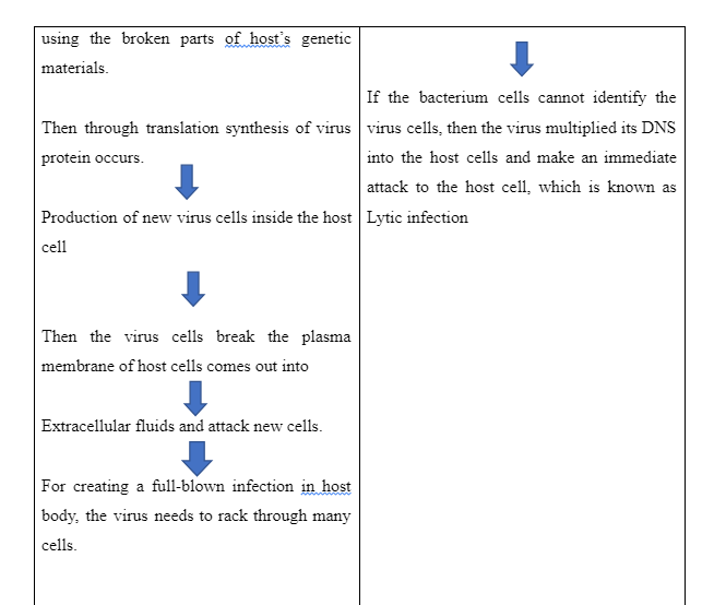
Contrast and compare the eukaryotic and prokaryotic cells and explain impact on viruses on them:
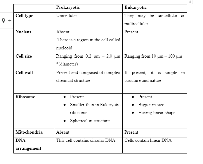
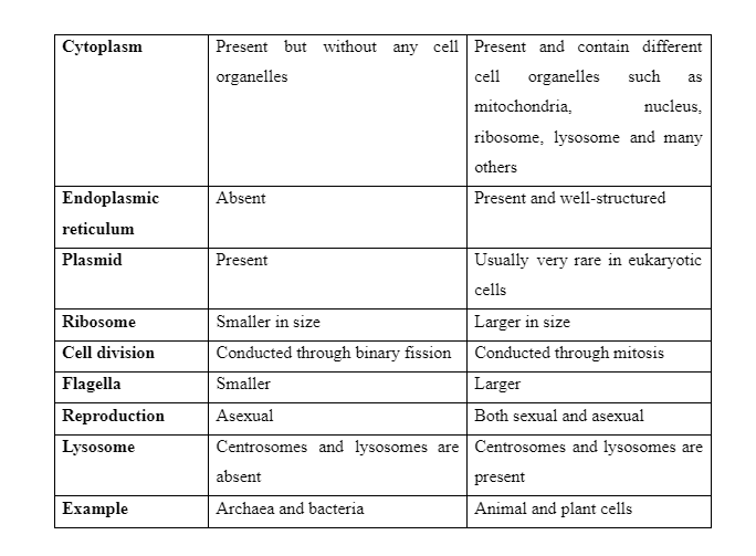

Discus eukaryotic subcellular structure and organelles:
As eukaryotic cell consists of two main structures such as cytoplasm and nucleus. In addition to these two structures there are also other membrane bound organelles such mitochondria, endoplasmic reticulum, Golgi bodies, plasmids, ribosome and lysosome (Gálvez-Iriqui et al. 2020).

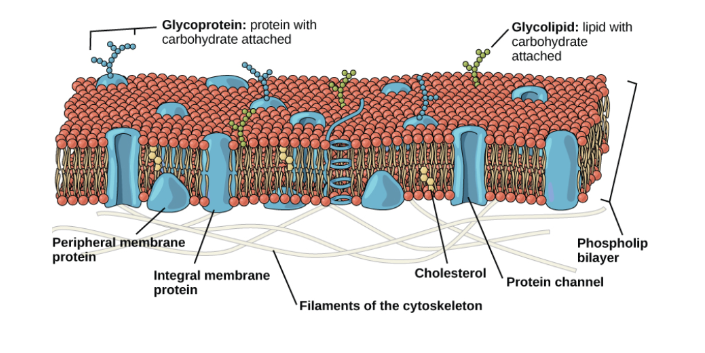
Plasma membrane:
Plasma membrane of a eukaryotic cell is formed of a phospholipid bilayer structure containing embedded proteins. Therese protein allows the plasma membrane to sperate its internal organism from the extracellular space in the outside the cell (Gálvez-Iriqui et al. 2020). Plasma membrane is composed mainly of proteins, lipid and water. Lipid forms the 40% structure plasma membrane. Lipids is formed if fatty acid ester and cholesterol. The backbone m of membrane lipids is made up of glycolipids, the three-carbon molecule. Lipid bilayer that surrounded the cells is amphipathic is nature with containing hydrophilic heads in one end and the hydrophobic end on the other end. For those seeking biology dissertation help, understanding these fundamental structures is essential for analysing cell membrane functions in greater detail.
Plasma membrane provides passage to the organism molecules such as ions, oxygen and water to come in and out of the cells. On the other hand, plasma membranes allow the cells to discharge the wastes materials out the cells through protein transported that carry these waste products and molecules and pass them out of the cells.
Cytoplasm:
Cytoplasm is referred to the region that is situated between the plasma membrane and nuclear membrane (Rout and Field, 2017). Cytoplasm of eukaryotic cells is consisting of a jelly like cytosol, cytoskeleton and some chemical. The organelles that are suspended into the jelly like cytosol are together cells as cytoskeleton. Evidences have shown that although cytoplasm is composed of 70-80% of water it does nit show semisolid texture. Other than cytoskeleton the chemicals that are present in the cytosol are proteins, polysaccharides, sugars, amino acids, nucleic acids and fatty acids.
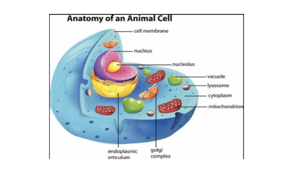
Nucleus:
Nucleus is one of the most crucial structure of a cells that consist of the genetic Materia (DNA). The membrane the surrounds nucleus is called nuclear envelop. It is phospholipid bilayers that form the inner and outer membrane. The membrane has pores that provides passage to exit and entry of ions, molecules and RNA between cytoplasm and nucleoplasm. Inside the nucleus there is the semisolid structure which is known as nucleoplasm. In the nucleoplasm there are nucleolus and chromatin. Nucleolus is the condensed region of chromatin in which ribosome is synthesized.
Ribosome:
ribosomes are attached to the cytoplasmic sides of the plasma membrane. These structures are associated with protein synthesis (Carreno et al. 2016). While viewing under the microscope, there are seen as clusters or single structures. Under microscopic view, they are look life tiny dots in that float freely on the cytoplasm.
Take a deeper dive into Specialized Tissue Generation> with our additional resources.
Mitochondria:
Mitochondria are known as the power house or energy factory of cells. This is because, mitochondria are the primary places in which metabolic respiration takes place. Depend on overall types of mitochondria, the process of respiration in aerobic and anaerobic (Røst et al. 2020). In case of aerobic respiration oxygen acts as the terminal electron. On the other hand, in case of anaerobic respiration, a compound other than oxygen acts as the terminal electron. In anaerobic respiration, ATP is produced through oxidative phosphorylation.
Mitochondria is the oval shaped structure that is surrounded by the phospholipid bilayer wit inner and outer membrane. On this mitochondrial membrane there many pores that contain protein reception that are involved in transferring ions, molecules inside and outside the mitochondria. The inner membrane of mitochondria has finger like projects which are known as cristae. The region that is located inside the cristae is known as Mitochondrial matrix.
Endoplasmic reticulum: (ER)
In eukaryotic cells there are interrelated compartments, that are collectively known as endoplasmic reticulum or ER (Carreno et al. 2016). These are the continuous membrane bounded structure a that is attached to the outer side of the nuclear envelope. ET is the convoluted membrane that created several delimited compartments that are interconnected through the fusion and fission of the membrane. The space that is coated in the endoplasmic reticulum is lumen.
Golgi apparatus:
There are the densely stained and reticular structure located near the nucleus. Camillo Golgi (1898) had first seen and discovered this structure in the cell, therefore the name, Golgi body is given the structure. Golgi body is compose on several disc like parallelly arranged sacs that are called cisternae. Each cisterna contain two sides one is the convex cis or forming side and the other is the concave trans side or maturing side.
Section 2:
The role of the cell membrane in regulating how nutrients are gained and waste products lost:
Plasma membrane develops selectively permeable barriers between the extracellular environment and the cell. Plasma membrane is composed of two layer of phospholipids that are selectively permeable. This permeability properly of plasma membrane assist cells to maintain balanced environment inside the cells (Carreno et al. 2016). Through regulating the entry of different molecules and nutrients into the cells and exit of waste from the cells to the extracellular matrix, plasma membrane allows cell to maintain the intrinsic property.
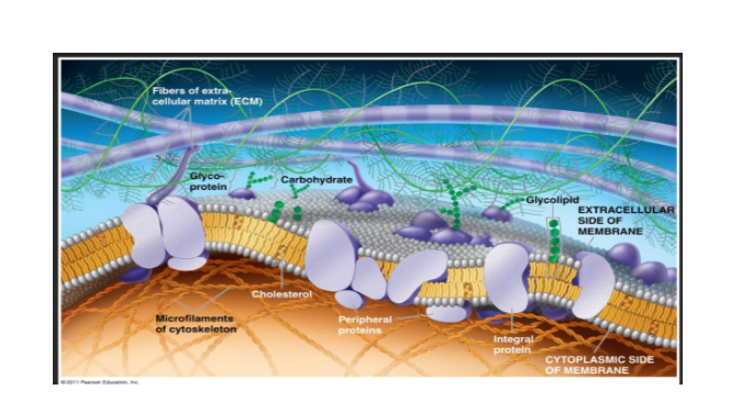
Plasma membrane are impermeable to the water-soluble molecules such as amino acids, glucose and ions. Therefore, the transportation of these molecules inside the cells occurs through the transport protein that are situated on the outer sides of the lipid bilayer ((Boiteux et al. 2017). Not all proteins carry all types of molecules. There are specific transport protein that carry specific molecules. Such as glycoprotein has protein and sugar moiety, thereby carrying only proteins and sugars into the cell. On the other hands, lipid proteins can only carry fat molecules.
Plasma membrane of different cell consist of specific sets of transport proteins that allows entry and exit of only specifics molecules. For example, the plasma membrane of epithelial cells consists of transport protein that transport only ions and small molecules inside the cells. Transport protein located on the plasma membrane on the intestine transport the digestion molecules such as amino acids and sugar into the blood cells.
Waste products such as ATP, CO2 and other lipid soluble molecules that are generated during the anaerobic or aerobic respiration inside the cells are transported outside the cell through simple diffusion (Rout and Field, 2017). In this process, the waste molecules are carried by the transporter protein from high concentration cytoplasm of call to the low concentration extra cellular spaces to maintain the equilibrium. Here the waste molecules are diffused from the cells to the outside space by flowing the physiochemical laws ad electrochemical gradients.
How animal cells use nutrients to provide the energy for growth, movement, and cell division
Animals need to take well-balanced diet that will assist the cells to get enough essential nutrients that are need to for sufficient energy reduction in terms of regulating different cell function such as cell division, growth and cell movements.
Organic molecules and energy that are required for building tissues and the cellular material come from foods. Sugar or carbohydrates are the major source of energy in animal cells. During the process of digestion, the carbohydrates are broken down into simple glucose molecules that provides the sufficient energy to tei cell through the metabolic pathway (Rout and Field, 2017). The excess glucose or sugar that are not used in energy production are then stored in the tissues of muscle and liver as glycogen through the process of glycogenesis. This stored glycogen is used to fuel while there is need to extra energy such a when animal runs.
Another important nutrient that anima need is organic nitrogen. Animal gets sufficient amount of nitrogen into their body trough consuming protein enriched foods. Nitrogen are the string source of amino acids that act as the building blocks of cell proteins. After consuming protein enriched food these proteins are broken down into amino acids that provided the good source of carbon and nitrogen into animal cells. Nitrogen and carbon act as the building blocks for nucleic acids, nucleotides, cells, proteins and tissues. Excess nitrogen that re not used in energy production are then exerted from the body as waste products such as urine.
Fats are also good source of energy and nutrients into the animal cells. Through consumed foods containing fats, metabolism occurs that breaks the fats into fatty acids. Through the metabolism, cells can get the fat-soluble vitamins and hormones that are important to regular normal function cells. The fatty that is produces due to fat metabolism provides good sources of phospholipids that are the major component of plasma membrane.
Essential nutrients:
There are some nutrients that cannot be formed by the animal bodies therefore animals required to receive these nutrients through foods. These nutrients are highly crucial in maintaining normal functions of cell, therefore they are called as essential nutrients (Carreno et al. 2016). Vitamins, minerals, fats, carbohydrate, water and proteins are six essential nutrients that animals can get through foods.
Minerals and different types of vitamins such as Vitamin A, D, E and B12 enter into animal body through foods. There are two categories of vitamins such as water soluble and fat-soluble vitamins. Fat soluble vitamins can easily be disabled in fats, thereby easily stress in muscles and tissues. On the otherhood water soluble vitamins is dissolved in water before their absorption into body. This is why the water-soluble vitamins can not be stored inside the body. Vitamins play important roles in animal body. For example, vitamin D are essential for developing strong bones and ligaments. On the other hand, Vitamin, A involved in eye development, strengthening immune system and supporting hair, nail and skin development.
Fat soluble vitamins:
These vitamins are generally found in foods containing oil and fats such as vegetable oil, animal fats, dairy floods and fatty acids animal cells need these vitamins on regular basis to perform their function smoothly (Freund et al. 2019). Animal body does not need these vitamins immediately, rather these vitamins are stored into the muscle and lever tissue for future use. Fat soluble vitamins are Vitamin, D, vitamin A, Vitamin K and Vitamin E.
Unlike the fat-soluble vitamins, the water-soluble vitamin cannot be stored in the body. After using the sufficient amount of these through metabolism for emery production, the excess vitamins come out of body through urination. Animal cells an get these vitamins through foods lie fruits, grain and vegetables.
The role of nucleic acids in the nucleus and cytoplasm:
Nucleic acids are the long chain polymeric macro molecules that are made up of several nucleotides (Gálvez-Iriqui et al. 2020). There are two major categories in nucleic acids in cells such as DNA [Deoxyribonucleic acid] and RNA [ribonucleic acid].
Function of DNA:
- DNA carries genetic information and transferred them from parent to offspring
- It controls and monitor the overall synthesis of RNA.
- Nitrogen base sequence in DNA determines which protein would be synthesized in new cell (Di Pierro et al. 2018)
- Double helical structure of DNA ensures no genetic disorders occurs in cells. In this context, the second identical strand of Deoxyribonucleic acid [DNA] runs anti-parallelly to the first and provides backup for lost and destroy of genetical information in the first strand (Rout and Field, 2017)
Function of RNA:
- RNA plays important roles in protein synthesis in new cells by using the transported genetical information (Wiseman et al. 2018)
- mRNA (messenger ribonucleic acid) plays an important role to transfer genetic information from parent to new cells through plasma membrane.
- Nucleic acids play important roles in mitosis and meiosis.
- Nucleus acid also assist in cellular respiration to provide energy to cell to perform its regular function/.
Process of protein synthesis:
This is the process in which the polypeptide chains are produced through using the combination of cedes of single amino acids within the cell. Protein synthesis is done in ribosome and nucleus of a cells (Røst et al. 2020). The entire process pf protein synthesise is mediated a controlled by the two major nucleus acids m, DNA and RNA.
Protein synthesis occurs in two steps such as:
- Transcription
- Translation
Transcription
Transcription is the process in which mRNA is formed from DNA by the involvement of RNA polymerase.
The process occurs in three steps.
- Initiation: The first step is known as initiation place, in which RNA polymerase binds to the promoter region of DNA strand by sigma bond thereby initiating transcription (MacGregor et al. 2019).
- Elongation: this is the second step of transcription in which RNA polymerase use the nucleotide triphosphate as the substrate thereby leads to polymerisation of the nucleotides of the templates as the complementary strands.
- Termination: In the last step, the termination place, a termination factor called, rho factors attach to the termination side of DNA strand thereby replacing initiation factors. In this stage DNA detaches from the RNA polymerase enzyme by rho factor (Shi et al. 2018).
- In prokaryotic cell the transcription occurs in the cytoplasm, whereas in the eukaryotic cell the process occurs in the nucleus.
Translation
- Translation is the second stage of protein synthesis that is occurred in ribosome.
- In this process, the messenger RNA [mRNA] contributes to the way to attach to the ribosome with help of ribosomal RNA and specific enzyme.
- tRNA or transfer RNA consists of single amino acid as well as the coded sequence which acts like the key. The key attached and fits to three code sequence of mRNA. Each set with three amino acid based is known as codon.
- Then the large subunit of ribosome attached to the small subunit. The full unit small and large subunit of ribosome, mRNA and tRNA are together called as initiation complex. The large subunit of ribosome consists of three suites such as A, P and E site.
- At this stage a tRNA attached to the A site of the large ribosome and then a bond between the amino acids and the methionine of the two tRNA molecule are formed (Won et al. 2016). Then the previous tRNA is deacylated then it is discharge from the ribosome through E site.
- Then the tRNA situated in A site of mRNA goes to the P site and a New tRNA carrying amino acid molecule to the A site of ribosome, repetition of this process is called elongation in which a strand of amino acids is formed.
- Finally, when the UAG codon which is known as stop codon teachers the ribosomal E site the elongation is terminated.
Section 3:
The generation of specialised tissues from embryonic stem cells.
- Embryonic stem cells or ES are the pluripotent cells that are developed from the interion cell mass of blastocyst during the pre-implantation period (MacGregor et al. 2019).
- ES have the ability to develop any types of cells expect placental cells. Only the ES of morula are totipotent cells that have the ability to differentiate into placental cells.
- Embryonic stem cells are differentiated into tissues of different cells such as blood cells, neurons, muscle and pancreatic cells
The process of interphase and factors that initiate cell division, and their importance:
Interphase:
Interphase is the stage in which cells prepares itself for cell division. In this stage cell synthesize protein, acquire nutrients and start DNS replication (Markov and Kaznacheev, 2016). The process of interphase is divided into three stages such as Gap 1, Synthesis and Gap 2 phase.
Gap 1:
After completion of division of chromosome, cell membrane of a cell is divided through cytokinesis and produces two cells these two cells then enter into gap 1 or G1 phase (Terzaghi et al. 2016). In this phase cell grow in size cells also replicate different organelles as per its requirement. Sometimes cells immediately leave the G1 phase and enter into the G0 phase. However, the cells that are most active never go the G0 phase rather they complete the entire interphase and then go to the cell division.
Synthesis:
This stage starts with production of two entwined DNA strand. In this stage the DNA polymerase enzyme leads to production of new strands to complete each pair of DNA. The two identical copy of chromise is are developed that are bound to each other forming sister chromatids. If the gametes are formed from parent cell then meiosis cell division is going to start after the interphase. On the other hand, if the cell is somatic cell then it will go for mitosis I which sister chromatids will be separated from each other and go to two new cells.
Gap2:
After the DNA replication is completed the cell enters this G2 phase. In this phase the cell volume of cytoplasm is increased (Elkouby and Mullins, 2017). Here cell undergoes replication of cell organelles such as mitochondria, ribosome and lysosome.
How the same genetic information is received by each daughter cell.
Through mitosis cell division two identical cells are developed from the parent cells. Two identical cells have the similar characteristics as the same genetic information are transferred through parent cells to daughter cells through mitosis process.
In this process there are five phases such as prophase, prometaphases, metaphase, anaphase, telophase.
During prophase chromatic are tightly coiled and twisted in which there are sister chromatids that are linked with centromere. In this stage the mitotic spindle is formed (Markov and Kaznacheev, 2016).
Each centromere contains radial arrays of microtubules or esters
Then nucleus breaks down and the centrosomes are located to the poles of the cell.
The chromosomes are lined up in the metaphase plate
The M check post ensure that sister chromatics are located to the opposite ends of mitotic spindle.
Then the anaphase begins and enzyme separase cleaves the cohesion that hold the sister chromatids together,
Two chromosomes are formed are the telophase and then cytokinesis occurs in which the plasma membrane of the cell divide the cytoplasm into two different cells each with one set of chromosomes.
The two daughter cells receive the identical characteristics from the parent cells as the chromosome that are developed from the parent chromosome are identical.
Dig deeper into Biology and Our Experiences with our selection of articles.
Reference list:
Appadurai, D., Gay, L., Moharir, A., Lang, M.J., Duncan, M.C., Schmidt, O., Teis, D., Vu, T.N., Silva, M., Jorgensen, E.M. and Babst, M., 2020. Plasma membrane tension regulates eisosome structure and function. Molecular Biology of the Cell, 31(4), pp.287-303.
Boiteux, S., Coste, F. and Castaing, B., 2017. Repair of 8-oxo-7, 8-dihydroguanine in prokaryotic and eukaryotic cells: Properties and biological roles of the Fpg and OGG1 DNA N-glycosylases. Free Radical Biology and Medicine, 107, pp.179-201.
Calderon, D., Nguyen, M.L., Mezger, A., Kathiria, A., Müller, F., Nguyen, V., Lescano, N., Wu, B., Trombetta, J., Ribado, J.V. and Knowles, D.A., 2019. Landscape of stimulation-responsive chromatin across diverse human immune cells. Nature genetics, pp.1-12.
Carreno, A., Gacitúa, M., Fuentes, J.A., Páez-Hernández, D., Peñaloza, J.P., Otero, C., Preite, M., Molins, E., Swords, W.B., Meyer, G.J. and Manriquez, J.M., 2016. Fluorescence probes for prokaryotic and eukaryotic cells using Re (CO) 3+ complexes with an electron withdrawing ancillary ligand. New Journal of Chemistry, 40(9), pp.7687-7700.
Chameettachal, A., Pillai, V.N., Ali, L.M., Pitchai, F.N.N., Ardah, M.T., Mustafa, F., Marquet, R. and Rizvi, T.A., 2018. Biochemical and Functional Characterization of Mouse Mammary Tumor Virus Full-Length Pr77Gag Expressed in Prokaryotic and Eukaryotic Cells. Viruses, 10(6), p.334.
Corces, M.R., Granja, J.M., Shams, S., Louie, B.H., Seoane, J.A., Zhou, W., Silva, T.C., Groeneveld, C., Wong, C.K., Cho, S.W. and Satpathy, A.T., 2018. The chromatin accessibility landscape of primary human cancers. Science, 362(6413).
Di Pierro, M., Cheng, R.R., Aiden, E.L., Wolynes, P.G. and Onuchic, J.N., 2017. De novo prediction of human chromosome structures: Epigenetic marking patterns encode genome architecture. Proceedings of the National Academy of Sciences, 114(46), pp.12126-12131.
Elkouby, Y.M. and Mullins, M.C., 2017. Coordination of cellular differentiation, polarity, mitosis and meiosis–new findings from early vertebrate oogenesis. Developmental biology, 430(2), pp.275-287.
Freund, I., Buhl, D.K., Boutin, S., Kotter, A., Pichot, F., Marchand, V., Vierbuchen, T., Heine, H., Motorin, Y., Helm, M. and Dalpke, A.H., 2019. 2′-O-methylation within prokaryotic and eukaryotic tRNA inhibits innate immune activation by endosomal Toll-like receptors but does not affect recognition of whole organisms. RNA, 25(7), pp.869-880.
Fullard, J.F., Hauberg, M.E., Bendl, J., Egervari, G., Cirnaru, M.D., Reach, S.M., Motl, J., Ehrlich, M.E., Hurd, Y.L. and Roussos, P., 2018. An atlas of chromatin accessibility in the adult human brain. Genome research, 28(8), pp.1243-1252.
Gálvez-Iriqui, A.C., García-Romo, J.S., Cortez-Rocha, M.O., Burgos-Hernández, A., Burboa-Zazueta, M.G., Luque-Alcaraz, A.G., Calderón-Santoyo, M., Argüelles-Monal, W.M. and Plascencia-Jatomea, M., 2020. Phytotoxicity, cytotoxicity, and in vivo antifungal efficacy of chitosan nanobiocomposites on prokaryotic and eukaryotic cells. Environmental Science and Pollution Research, pp.1-15.
Grubert, F., Srivas, R., Spacek, D.V., Kasowski, M., Ruiz-Velasco, M., Sinnott-Armstrong, N., Greenside, P., Narasimha, A., Liu, Q., Geller, B. and Sanghi, A., 2020. Landscape of cohesin-mediated chromatin loops in the human genome. Nature, 583(7818), pp.737-743.
Jacobson, K., Liu, P. and Lagerholm, B.C., 2019. The lateral organization and mobility of plasma membrane components. Cell, 177(4), pp.806-819.
Jain, M., Olsen, H.E., Turner, D.J., Stoddart, D., Bulazel, K.V., Paten, B., Haussler, D., Willard, H.F., Akeson, M. and Miga, K.H., 2018. Linear assembly of a human centromere on the Y chromosome. Nature biotechnology, 36(4), pp.321-323.
Jobling, M.A. and Tyler-Smith, C., 2017. Human Y-chromosome variation in the genome-sequencing era. Nature Reviews Genetics, 18(8), p.485.
Lee, S.H., Kim, Y.J., Ho Kang, B. and Park, C.K., 2019. Effect of nicotinic acid on the plasma membrane function and polyunsaturated fatty acids composition during cryopreservation in boar sperm. Reproduction in Domestic Animals, 54(9), pp.1251-1257.
Lorent, J.H., Diaz-Rohrer, B., Lin, X., Spring, K., Gorfe, A.A., Levental, K.R. and Levental, I., 2017. Structural determinants and functional consequences of protein affinity for membrane rafts. Nature communications, 8(1), pp.1-10.
MacGregor, I.A., Adams, I.R. and Gilbert, N., 2019. Large-scale chromatin organisation in interphase, mitosis and meiosis. Biochemical Journal, 476(15), pp.2141-2156.
Markov, A.V. and Kaznacheev, I.S., 2016. Evolutionary consequences of polyploidy in prokaryotes and the origin of mitosis and meiosis. Biology direct, 11(1), p.28.
Nakanishi, H., Irie, K., Segawa, K., Hasegawa, K., Fujiyoshi, Y., Nagata, S. and Abe, K., 2020. Crystal structure of a human plasma membrane phospholipid flippase. Journal of Biological Chemistry, pp.jbc-RA120.
Partridge, E.C., Chhetri, S.B., Prokop, J.W., Ramaker, R.C., Jansen, C.S., Goh, S.T., Mackiewicz, M., Newberry, K.M., Brandsmeier, L.A., Meadows, S.K. and Messer, C.L., 2020. Occupancy maps of 208 chromatin-associated proteins in one human cell type. Nature, 583(7818), pp.720-728.
Røst, L.M., Brekke Thorfinnsdottir, L., Kumar, K., Fuchino, K., Eide Langørgen, I., Bartosova, Z., Kristiansen, K.A. and Bruheim, P., 2020. Absolute quantification of the central carbon metabolome in eight commonly applied prokaryotic and eukaryotic model systems. Metabolites, 10(2), p.74.
Rout, M.P. and Field, M.C., 2017. The evolution of organellar coat complexes and organization of the eukaryotic cell. Annual review of biochemistry, 86, pp.637-657.
Shan, L., Wu, C., Chen, D., Hou, L., Li, X., Wang, L., Chu, X., Hou, Y. and Wang, Z., 2017. Regulators of alternative polyadenylation operate at the transition from mitosis to meiosis. Journal of Genetics and Genomics, 44(2), pp.95-106.
Shi, G., Liu, L., Hyeon, C. and Thirumalai, D., 2018. Interphase human chromosome exhibits out of equilibrium glassy dynamics. Nature communications, 9(1), pp.1-13.
Swanson, G.M., Estill, M., Diamond, M.P., Legro, R.S., Coutifaris, C., Barnhart, K.T., Huang, H., Hansen, K.R., Trussell, J.C., Coward, R.M. and Zhang, H., 2020. Human chromatin remodeler cofactor, RNA interactor, eraser and writer sperm RNAs responding to obesity. Epigenetics, 15(1-2), pp.32-46.
Terzaghi, L., Tessaro, I., Raucci, F., Merico, V., Mazzini, G., Garagna, S., Zuccotti, M., Franciosi, F. and Lodde, V., 2016. PGRMC1 participates in late events of bovine granulosa cells mitosis and oocyte meiosis. Cell Cycle, 15(15), pp.2019-2032.
Wiseman, F.K., Pulford, L.J., Barkus, C., Liao, F., Portelius, E., Webb, R., Chávez-Gutiérrez, L., Cleverley, K., Noy, S., Sheppard, O. and Collins, T., 2018. Trisomy of human chromosome 21 enhances amyloid-β deposition independently of an extra copy of APP. Brain, 141(8), pp.2457-2474.
Won, H., de La Torre-Ubieta, L., Stein, J.L., Parikshak, N.N., Huang, J., Opland, C.K., Gandal, M.J., Sutton, G.J., Hormozdiari, F., Lu, D. and Lee, C., 2016. Chromosome conformation elucidates regulatory relationships in developing human brain. Nature, 538(7626), pp.523-527.


- 24/7 Customer Support
- 100% Customer Satisfaction
- No Privacy Violation
- Quick Services
- Subject Experts