Cardiac Control/Ecg Scenario
Question A
- Sino atrial node (SAN)-These are neurons found in the right atrium of the heart on the upper part. They act as the heart’s pacemaker initiates the cardiac cycle.
- Atrio ventricular node (AVN)-This is a group of cardiac muscles which are specialized fibers located in the central part of the heart. This node takes signals from the SA node, it slows them down and then regulates them to prevent atrial fibrillation which is rapid conduction. This also ensures that the atria are empty and closed before it stimulates the ventricles. It sends electrical impulses to the ventricles from the atria (Manfredi, 2014). For students working on related research, seeking healthcare dissertation help can provide valuable insights and guidance in understanding complex topics like AVN.
- Bundle of His and Purkyje fibres-This is an important part of the electrical conduction system of the heart. These are fibers specialized in transmitting impulses from the atrioventricular node to the heart ventricles. They distribute impulses to the ventricular muscles (Manfredi, 2014).
b)

CARDIAC CONTROL/ECG SCENARIO

- P wave-This is the first wave on an ECG showing an atrial systole. This is atrial depolarization whereby the atria contracts. Decreased or increased P waves show a problem with the concentration of potassium ion in the body which will alter nerve activity. Missing P wave shows atrial fibrillation- a cardiac arrhythmia where the heart beats irregularly, resulting to an inefficient ventricular diastole (Lee & Chen, 2007).
- QRS wave-a combination of Q, R, and the S waves. It shows ventricular systole- ventricular depolarization and contraction. It indicates action potentials traveling from the AV node, into the ventricular muscle tissue via the bundle of His and Purkinje fibers. QRS complex abnormalities can indicate myocardial infarctions or cardiac hypertrophy
- T wave shows ventricular repolarization where by following depolarization and contraction the ventricles relax. The gap between the T wave and the S wave is known as the segment. It indicates the time in between a ventricular depolarization and repolarization. An increased ST segment indicates myocardial infarctions and a decreased or missing ST segments may be an indication of myocardial ischemia (Lee & Chen, 2007).
c)
a. Calculating the heart rate for each trace.
The calculation is based on the entire ECG being 10 seconds. The number of beats or the heart rate per minute is calculated by counting the number of QRS complexes the multiply them by six since 10 seconds (for an entire ECG )multiplied by 6 equals to 60 seconds which is 1 minute.

- 12×6=72 beats per minute
- 7×6=42 beats per minute
- 5×6=30 beats per minute
- 6×6=36 beats per minute
b)
- Condition- Bradycardia
- Condition-Tachycardia
- Condition- Sinus rhythm
- Condition- Atrial Fibrillation
c)

During an exercise, body muscles become active, and therefore they respire fast causing various changes to the blood. Some of these changes are reduced the oxygen concentration in the blood, elevated carbon dioxide and decreased pH and also increased temperatures. Carbon dioxide in high amount is produced which dissolves in the blood to form carbonic acid thus increase the acidity in the blood lowering the pH. The changes are detected by different receptor cells all over the body (Manfredi, 2014). The most sensitive change is the pH variations and hence it is the most important. The major receptor cells (chemoreceptors) that detect chemical changes are located in the following areas:
- The walls of the Aorta -Has the aortic body which monitors blood as it leaves the heart. The carotid bodies on walls of the carotid arteries controls blood moving to the head and the brain (Manfredi, 2014).
- The medulla-which monitors the tissue fluid found in the brain. These chemoreceptors detect the chemical changes; they then send nerve impulses to the cardiovascular centers showing that high respiration is occurring. In response to this, the cardiovascular center increases the heart rate.
 Exercise also affects the rest of circulation and also increases cardiac output. During an exercise, respiration rate is very high, and therefore the body requires large amounts of oxygen. The heart is forced to increase its production to increase the amount of blood flowing in the body through the capillaries at the muscles (Manfredi, 2014).
d) ECG Case Study
- A cardiac dysrhythmia can be described as an abnormal heart beat. It may be either low or high, but it may be irregular in its passing. Some cardiac dysrhythmia is life-threatening while others are not. From the above ECG trace, the heart rhythm is very slow (below 60 beats per minute).This is cardiac dysrhythmias know as Brady arrhythmias.
- If one has high blood pressure, it implies that their heart has to overwork to pump blood all over the body. To cope with this the core muscles especially at the left side of the heart become thicker and stiffer making the heart become enlarged. The left side pumps blood to all part, and therefore it is the most affected as to all regions of the body. The left ventricles hence becomes enlarged, and this is known as left ventricular hypertrophy (LVH)
- The left and the right side of the heart perform different tasks. The right side receives blood that has already circulated in the body parts and pumps it to the lungs. Therefore it is not affected when the blood pressure is high as it is not associated with getting blood around the body (Manfredi, 2014).
- Ramipril belongs to a group of drugs called ACE (angiotensin-converting enzyme) inhibitors which relax the muscles around the arterioles. They make the arterioles expand, and thus more blood can flow through more easily.This increases cardiac output. Digoxin drugs belong to a group of medicines known as cardiac glycosides. It functions by affecting specific minerals inside the heart cells such as sodium and potassium. It helps reducing pressure in the heart and assists in maintaining a healthy, regular and strong heartbeat. It leads to increased cardiac output (Manfredi, 2014).
- Caffeine binds directly with vascular smooth muscle cell receptors causing vasodilatation. It also blocks the adenosine receptors which are essential in coronary arteries to help in coronary blood flow during exercise. It also affects all other adenosine receptors, and this leads to a compensatory increase in adenosine in the body. This stimulates chemoreceptors used in circulation and any other receptor, and this leads to increase in sympathetic node among other effects resulting in increased blood pressure.
Part B - Oedema (Criteria 4.1 and 4.2)

A
Circulation of blood is from the heart through the arteries and then to the capillaries. The blood in the arteries and arterioles is at high pressure, and it enters the capillaries still at high pressure. Hydrostatic pressure forces blood fluid through the thin capillary walls to the surrounding tissues, and this leads to filtration of the fluid (Melenovsky & Hwang, 2014). This plasma is known as the tissue fluid. This movement of water dissolved solutes apart from proteins through the thin wall of the capillaries occurs through the process of diffusion and filtration. Only dissolved solutes and small molecules move through the capillary walls. Substances like blood cells do not pass through since they are large and they remain in the blood fluid.
Dissolved substances that are small enough and water make up the tissue fluid since they are small enough to pass through the capillaries. It’s not only the hydrostatic pressure of the blood that enables this process, but there are still other forces. The fluid also has its hydrostatic pressure that pushes it back to the blood and hence it cannot accumulate in the tissue spaces. Osmosis is involved in the movement of water from the fluid to blood. This is the process through which fluids and dissolved substances in them pass through a membrane till a balance in all substances involved is reached a balance. The tissue and the blood fluids have dissolved substances, and that gives them a negative water potential. However, the tissue fluid has less water potential as compared to the blood fluid, and therefore water from the tissue fluid flows back to the blood through osmosis. The excess fluid that is not absorbed back to the blood capillaries drains into the lymphatic system via the lymphatic vessels where it later combines with blood (Melenovsky & Hwang, 2014).
The lymphatic system consists of a network of vessels, and it takes nutrients to cells and takes waste products from them. It has lymph vessels and lymph capillaries which are somehow similar to the blood capillaries and blood vessels. It also has lymph ducts and lymph nodes. Lymph ducts are tube-like structures that carry fluid from the secreting glands. Lymph nodes filter out toxins and bacteria that may be present in the lymph passing through them. The lymph is similar in composition with the tissue fluid, but it has less oxygen concentration and very few nutrients and some fatty acids absorbed from the intestines.
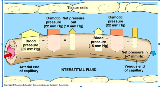
Figure 3
B
Edema is the accumulation of fluids in the body tissues. The conditions below are just some of the conditions that may cause edema as they disrupt the normal flow of tissue fluid.
- Kwashiorkor-this is a disorder caused by lack of sufficient plasma proteins in the body as a result of a deficiency of proteins. Low levels of proteins in the body as a result of malnutrition, liver or kidney diseases can lead to edema. Proteins help in holding salts and water inside the blood vessels so that fluids do not leak out into the body tissues. The most common cause of malnutrition-related edema is protein since albumin which is a protein is the largest component of the blood (Abbas & Lichtman, 2014). Enough Albumins in the blood helps the body maintain balance in fluids and hence no excess fluid that can accumulate in the cells. In the case of kwashiorkor the levels of proteins in the body are reduced and as the body tries to maintain balance in concentrations fluids then leak out of the vessels, and this causes edema in the tissue spaces.

Right-sided heart failure
After blood has circulated through out the body it goes to the right side of the heart through the veins which pumps it to the lungs. A failure on the right side of the heart leads to reduced output from the right ventricles and blood flows back into the veins in the lower body and also the legs. This may result to pitting edema which a swelling that occurs in the tissues under the skin in the lower legs and feet. If the right side fails and loses pumping power blood accumulates in the body veins. This causes swellings in the legs and ankles and also swelling of the abdomen.
Elephantiasis
Elephantiasis is a disease which is caused by filarial worms which are parasitic worms. This disease is transmitted by female mosquito. Elephantiasis causes extreme swelling of the legs and arms. When the Anopheles mosquito carrying the disease bites a human being, it injects the larvae into the body. Then the small larvae move to the lymph glands and then to the lymphatic system where it develops into an adult worm. This worm restricts the normal flow of lymphatic fluid leading to swelling, discoloration, and thickening of the skin. That is what can lead to an elephant’s leg appearance.
Looking for further insights on Can gender have an impact on how people living with HIV? Click here.
References
Abbas, A. K., Lichtman, A. H., & Pillai, S. (2014). Cellular and molecular immunology. Elsevier Health Sciences.
Dabaghyan, M., Zhang, S. H., Ward, J., Kwong, R. Y., Stevenson, W. G., Watkins, R. D., ... & Schmidt, E. J. (2016). Automated removal of gradient-induced voltages from 12-lead ECG traces during high-gradient duty-cycle MRI sequences. Journal of Cardiovascular Magnetic Resonancem.
Lee, R. G., Chen, K. C., Hsiao, C. C., & Tseng, C. L. (2007). A mobile care system with alert mechanism. IEEE Transactions on Information Technology in Biomedicine.
Manfredi, S. (2014). Congestion control for differentiated healthcare service delivery in emerging heterogeneous wireless body area networks. IEEE Wireless Communications, 21(2), 81-90.
Melenovsky, V., Hwang, S. J., Lin, G., Redfield, M. M., & Borlaug, B. A. (2014). Right heart dysfunction in heart failure with preserved ejection fraction. European heart journal, 35(48), 3452-3462.


- 24/7 Customer Support
- 100% Customer Satisfaction
- No Privacy Violation
- Quick Services
- Subject Experts