EEG recording – Challenge
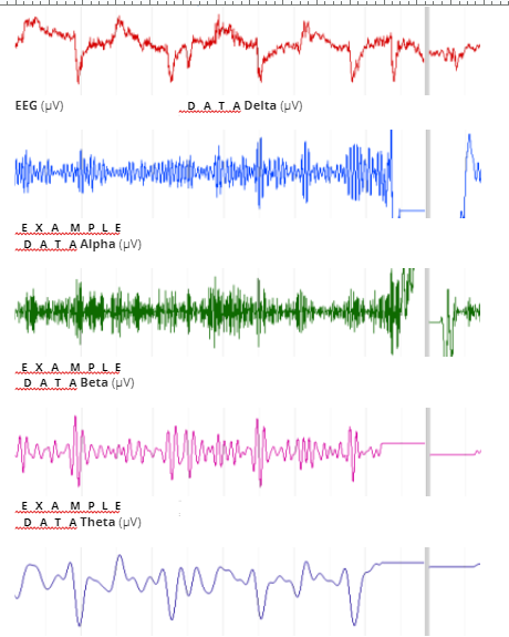
Q1) EEGs contain component "brain waves" of dierent frequencies. Position each label in the correct location. 4 pts

Alpha (8–13 Hz)
Beta (13–30 Hz)
Delta (0.5–4 Hz)
Theta (4–8 Hz)
Q2) EEG is a result of which of the following? 2 pts
Action potentials in the CNS.
Predominantly the visual cortex.
Slow changes in cortical neuron membrane potentials.
Q3) Which description most accurately denes EEG frequency and amplitude?
Frequency is measured in Hertz (Hz) and is dened as the number of times a wave oscillates per second. The amplitude is the time of the waveform measured from 0 seconds. Amplitude is measured in macro-volts (µV).
Frequency is measured in Hertz (Hz) and is dened as the number of times a wave peaks per second. Frequency is also known as a measure of EEG power. The amplitude is the height or peak measurement of the cycle measured from time 0 seconds. Amplitude is measured in macro-volts (µV) and reects EEG control.
Frequency is measured in Hertz (Hz) and is dened as the number of times a wave peaks per second. The amplitude is the circumference of the waveform as measured from the baseline. Amplitude is measured in micro-volts (µV) and typically divided into alpha, beta, gamma and theta.

Frequency is measured in Hertz (Hz) and is dened as the number of times a wave peaks per second. The amplitude is the height or peak measurement of the cycle as measured from the baseline. Amplitude is measured in micro-volts (µV) and also known as power.
All of the above
Recognizing artifacts – Activity
Alpha and beta rhythm – Activity

Alpha and beta rhythm – Analysis
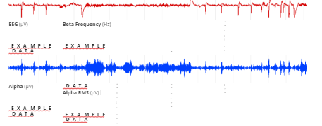
Q4) What happened to the alpha and beta waves when the eyes were closed compared to when they were 5 pts open? Explain why this happened [100-200 words]
alpha waves occur while the eyes are closed. That is because they disappear during sleep and vanish when the concentration is focused on one specic task. The alpha waves` rhythm could have a frequency between 8 and 13 Hz. During attention to stimuli and tasks, beta waves with higher frequency replace the alpha waves. Beta waves are quite common when one is concentrating on a task or when one is stressed or experiencing some sort of psychological tension. With eyes open or when there is some mental activity going on, alpha waves are observed to attenuate or decrease and are subsequently replaced by beta waves. The rhythm of beta waves is usually between 13 and 40 Hz. For those needing healthcare dissertation help, understanding these brain wave patterns can be crucial in exploring how mental states affect cognitive functions and overall health.
Q6) What eects did the mental arithmetic have on the alpha and beta wave activity? [100-200 words] 5 pts
The alpha waves amplitude, a representative component of the awake brain wave decreased by about 15-20% when the mental arithmetic was being conducted. This kind of amplitude suppression corresponds to an increase in the alpha waves temporal uctuation by between 15-30%. Particularly, the alpha wave`s amplitude reduction rate was up to 25% in the low-frequency range under mental arithmetic. On the other hand, the beta wave`s amplitude did not show any signicant change under the majority of the stress conditions, including even combined stress. From this observation, it is clear that mental arithmetic has an eect on the stabilization of the alpha wave in the low-frequency range, and this could be crucial to the measurement of mental stress through the use of brain waves.
Q7) Are alpha oscillations related to attention? Support your answer with empirical evidence [100-200 words] 5 pts
In the EEG, alpha oscillations have associations with attention and internal imagery. Without a doubt, alpha oscillations are the most noticeable features of EEG signals and they are dominant when an individual`s eyes are closed and also pop up when some eyes are also open. Attention is the brain's mechanism to focus on what is deemed as important, while concurrently suppressing any forms of potentially distracting and irrelevant information. Wolfgang Klimsech and Ole Jensen suggest that alpha synchronization increments are involved in suppressing information that is not relevant to tasks at hand, while an increase in the resynchronization of alpha oscillation releases inhibition which is instrumental for the facilitation of task-relevant processing.
Q8) Briey dene each of Gamma, Theta and Beta frequency bands and list one cognitive process associated 6 pts with each frequency band (provide references to support your answers) [1-2 sentences per frequency band]
The gamma-activity band is made up of an EEG frequency range from 30 to 200 Hz and is widely distributed throughout cerebral structures. GBA is involved in dierent cerebral functions, including attention, perception, consciousness, memory, motor control, and synaptic plasticity.
The theta rhythm, that is, the regular sinusoidal oscillations that happen in the frequency range of 4 to 7 Hz recorded in the hippocampal EEG are a prominent REM sleep signature in mammals, and this includes mammals.
The beta rhythms come in rvnges: beta 1 (13-20 Hz), beta 2 (21-30 Hz), and gamma (30-60 Hz)
Q9) What are the advantages and disadvantages of using EEG/ERP compared to other neuroscientic 5 pts techniques such as fMRI, PET and TMS? [100-200 words]
The ability to see the brain activity as it unfolds in real-time, even at the level of milliseconds is one of the biggest advantages of EEG/ERP. EEG is also relatively non-invasive and is, therefore, usable in human subjects.
In addition, the temporal resolution of EEG is relatively excellent, low cost, and does not have any real restrictions. Its spatial resolution is, however, poor and makes a lot of noise. However, with EEG, it is not easy to gure out the exact place in the brain where electrical activity originates from. When very many electrodes are put over the scalp, it becomes possible to know the points where ERP components are strongest. It is worth noting that this does not exactly point to where the brain signals come from but is useful in showing whether the two ERP components either come from the same place or from dierent places.
Q10) Dene "frontal EEG asymmetry" and critically evaluate the relationship with individual dierences in either depression OR anxiety. Make sure that you cite and reference all sources. [500 words]
Frontal EEG asymmetry is the dierence between left and right alpha activity over the brain's frontal regions during the recording of electroencephalographic (EEG). This has been researched widely in studies of psychopathology, motivation, and emotion, and is still a metric that has been quantied and analyzed through the use of procedures that are relatively diverse. Frontal EEG asymmetry is recognized as a rather promising neurophysiological marker of the risk of depression. It comes in handy in the prediction of negative eects and emotional responses (Smit, Posthuma, and Boomsma, 2007).
The Frontal EEG asymmetry model pioneered by Davidson (1983,1993) is supportive of the view that the brain systems activities act as moderators of motivational trait tendencies for approaching and withdrawing novel emotional stimuli and additionally mediating either the approach of withdrawal of those motivational tendencies that underlie emotion. In accordance with this model, an increase in the left prefrontal activity, whether as a state or a trait has associations with approach-related emotions, while increments in the activity in the right prefrontal have associations with withdrawal-related emotions. Diculties in releasing attention from negative stimuli have been found to be reliant on the low activity at the left frontal, the same case with anxiety arousal and depression (Coan and Allen, 2003). On the other hand, diculty in the inhibition of positive distractions has been found to be reliant on the low activity at the right frontal, with the same happening in addiction and poor self-regulation. This points to hypoactivation of the left frontal region being a representative marker for mood disorders.

Looking for further insights on Effects of Obesity on VO2 Peak: A Comprehensive Study? Click here.
Depression and anxiety are emotion-related disturbances that have links with relative right-sided resting frontal EEG asymmetry among adults and also infants whose mothers are aicted. Thibodeau, Jorgensen, and Kim (2006), report that symmetry is generated by the induced euphoria mood state, while the induced depression mood state has associations with the greater activation of the right frontal lobe. Stewart, Bismark, and Towers (2010), posit that the asymmetry of subjects acts as a predictor of the levels of negative aect as responses to negative lm clips, which has associations with enhanced activation of the right hemisphere. Rofriguez et al. (2015), also reports on the presence of signicant activations in various right frontal regions as a result of the induction of negative mood in the control group but not in expressive suppression and cognitive reappraisal groups. Gale et al. (2001), however, in an earlier study found out that negative mood leads to greater activation of the left frontal region, while the personalities of individuals mediate the direction of the right hemisphere`s dierentiation between negative and positive mood. The ndings of Warden-Smith et al. (2017), are perhaps the most interesting as they show that positive mood inductions yield a decrease of the asymmetry of the right frontal and an increase of left frontal asymmetry in a negative alpha fontal group. This is as though the change in alpha-wave activity in the positive aect`s direction happened in people who are susceptible to negative aect.
Continue your exploration of Education Beyond Content Knowledge for Holistic Development with our related content.
Reference list [add any references you have cited in this report below]
Coan, J.A. and Allen, J.J., 2003. The state and trait nature of frontal EEG asymmetry in emotion.
Davidson, R.J., 2004. What does the prefrontal cortex “do” in aect: perspectives on frontal EEG asymmetry research. Biological psychology, 67(1-2), pp.219-234.
Gale, A., Edwards, J., Morris, P., Moore, R. and Forrester, D., 2001. Extraversion–introversion, neuroticism–stability, and EEG indicators of positive and negative empathic mood. Personality and Individual Dierences, 30(3), pp.449-461.
Smit, D.J.A., Posthuma, D., Boomsma, D.I. and De Geus, E.J.C., 2007. The relation between frontal EEG asymmetry and the risk for anxiety and depression. Biological psychology, 74(1), pp.26-33.
Stewart, J.L., Bismark, A.W., Towers, D.N., Coan, J.A. and Allen, J.J., 2010. Resting frontal EEG asymmetry as an endophenotype for depression risk: sex-specic patterns of frontal brain asymmetry. Journal of abnormal psychology, 119(3), p.502.
Thibodeau, R., Jorgensen, R.S. and Kim, S., 2006. Depression, anxiety, and resting frontal EEG asymmetry: a meta analytic review. Journal of abnormal psychology, 115(4), p.715.
Warden-Smith, J., Paul, L., Olukogbon, K., Bointon, E.S., Cole, R.H., John, S.R., Dong, S. and Jacob, T.J., 2017. Light and smell stimulus protocol reduced negative frontal EEG asymmetry and improved mood. Open Life Sciences, 12(1), pp.51- 61.


- 24/7 Customer Support
- 100% Customer Satisfaction
- No Privacy Violation
- Quick Services
- Subject Experts