Understanding Human Muscle and Skeletal Systems
- 16 Pages
- Published On: 11-12-2023
Introduction
In this study, the anatomy and organisation of muscle and skeletal system on the human body is to be disused along with their functions. Thereafter, their mechanism of movement and way body repair broken bones is to be explained.
AC 11.1
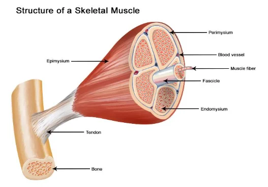
The muscle cells common called monocytes are the cells that contribute to the development of muscle tissues. There are mainly three distinct types of muscle tissues in the human body based on their structural orientation which are skeletal, smooth and cardiac muscles. The skeletal muscles are composed of many skeletal muscle fibres that consist of a single cylindrical muscle cell and they are bundled and wrapped together with connective tissue covering to form tuff of muscles (Ostrovidov et al., 2019). The connective tissue covering the muscles is known as epimysium and connective tissue over the epimysium is known as fascia that separates each of the muscles from another. The epimysium gets projected inside to form compartment for muscles and each of these compartments are composed of muscle fibres gathered in a bundle. A single bundle of skeletal muscle fibre is known as fasciculus and it is surrounded by perimysium. In the fasciculus, each of the muscle fibre is surrounded by endomysium (Dave et al., 2020). For students pursuing studies in this field, seeking psychology dissertation help can provide valuable insights into these intricate processes.

Continue your journey with our comprehensive guide to Impact on the Respiratory System.
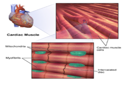
The cardiac muscle cells are present only in the heart and their structure shows they have muscle fibres with single nucleus in them. The cardiac muscle cells are branched and joined with each other through intercalated discs which have gap junctions in them to allow depolarisation of the cells and desmosomes so that muscles fibres are held together during the contraction of the heart (Litviňuková et al., 2020).

One of the basic animal tissues is connective tissue which develops from the mesoderm. The connective tissue composed of three parts which are cells that are suspended in the matrix or ground substance, fibres and cells. The ground substance or matrix is viscous and colourless fluid that fills the gaps present between the muscle fibres and cells. It is made up of proteoglycans along with cell adhesion proteins which help the connective tissues act as glue to cover the muscle fibres. The fibres in the connective tissue are mainly three types which are elastic fibres, collagen fibres and reticular fibres (Nishiguchi et al., 2019). The collagen fibres developed from fibrous protein are secreted in the extracellular space of muscles to provide increased tensile strength. The reticular fibres are fine and short collagenous fibres which for delicate framework through branching and the elastic fibres are elongated and thin fibbers which assist the connective tissue to get recoiled and stretched (Sefton and Kardon, 2019). There are mainly three types of connective tissue which are loose connective tissue, specialised connective tissue and dense connective tissue (Nishiguchi et al., 2019).


AC 11.2
The contraction of straited muscle is explained through the Sliding Filament theory. According to the theory, the muscle cell to contract requires its sarcomere to be shortened. However, the thin (actin) and thicker (myosine) muscle filaments of which the sarcomeres are composed shows inability to shorten and instead they show sliding movement over one another to shorten the sarcomere while keeping the filaments to remain of equal length. The mechanism of straited muscle contraction is bonding between the actin to myosine filament in forming the cross-linked bridge to promote movement of filament (Rassier, 2017). The sarcomere is the distance present between the two Z-discs of consecutive nature. During contraction of muscle, the distance between Z discs is reduced and the H zone which is the central region of the A zone containing only thick filaments are shortened. The I band containing the actin filaments also gets shortened. However, the A bands does not shorten and instead they move closer to one another during the muscle contraction eventually disappearing from the space. The actin is then pulled by the myosin towrads the sarcomere centre till the Z discs approaches the myosin filaments. The overlapping zone gets gradually increases with the actin filaments sliding inward due to support muscle movement (Smith, 2018).

AC 21.1
In the body, the skeletal system is made up of bones, cartilage, ligaments and other tissues that supports performing essential body functions. The bone which makes up the structure of the skeleton is developed of osseous tissues which are dense and hard connective tissue. In the body area such as joints where whole bones moves, cartilaginous tissues are present which are flexible, semi-rigid and smooth connective tissue to support the movement. The ligaments are made up of dense connective tissue which surrounds the area of the joint to ensure the skeletal elements remained joined in performing movement (Watson and Adams, 2018). The skeletal system is composed of four key fibrous and mineralised connective tissues such as bones, ligaments, tendons and joints (Watson and Adams, 2018).
The key types of bones in the skeletal system are long, flat, irregular, short and sesamoid. The flat bones which are present in the skull, thoracic cage and pelvis function in forming shield to protect the major internal organs of the body. The long bones such as the femur and small bones such as phalanges support to facilitate movement and managing weight of the body to stand. The short bones located in the writ and ankle support joint movement. The irregular bones which are different in shape are present in the vertebrae, pelvis and other supporting internal structure whereas the sesamoid bones that are embedded in tendons protects the tendon from wear and tear due to stress (Caon, 2020). The three main joints in the skeletal system are cartilaginous, fibrous and synovial joints. The fibrous joints are those which are connected by dense connective tissue rich in collagen fibres. The cartilaginous joints are bones forming joints supported by cartilage and found mostly in the vertebrae of the body. The synovial joints contain synovial cavity which are fluid-filled, in turn, acting as cushion for the end of the bone to move without stress. On the basis of the movement of synovial joints, they are named differentially (Caon, 2020; Popov et al., 2021).


AC 31.1
Continue your exploration of Human Behaviour with our related content.
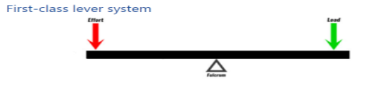
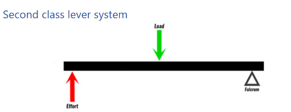
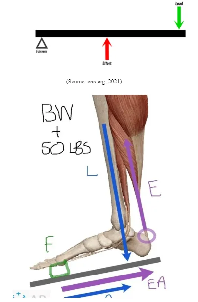
There are mainly three types of biomechanism of levers when the muscle forces transmit through the skeletal system which are first-class lever, second-class lever and third-class lever (cnx.org, 2021). In first-class lever system, the fulcrum is present in middle with equal distance from the effort and load arm. The effort arm is the distance between effort (muscle insertion site) and fulcrum (joint). The load arm is the distance between load (loaded body part) and fulcrum (joint). The biomechanism mentions the higher the ratio between the effort and load arm, more efficient the lever system (Mohamed et al., 2020). One example of first-class lever is the triceps are considered as the effort, elbow joint is the fulcrum and load on the arm is the load during javelin throw (cnx.org, 2021). The second-class lever is the biomechanism in which load is present in the middle with the fulcrum and effort lying on opposite side with equal distance from the load in the middle (cnx.org, 2021). The second-class lever example is exercise that involves planar flex at the ankle joint where the ball of the toes and foot acts as fulcrum, the body weight is the loaf and the gastrocnemius muscle is the effort (Cox, 2017). The third-class lever is the one in which the effort is present in the middle with load and fulcrum on the either end in equal distance from the effort (Wilke and Volkheimer, 2018). In most body function, the third-class lever biomechanism is used and one such example in movement of joints during running, jumping and other. In this condition during the knee flexion, the insertion of tibia hamstrings in the effort, the knee joint is the fulcrum and the leg is the load (cnx.org, 2021).

AC 31.2
The long bones grow continuously in length till adolescent with bone tissue being added at the epiphyseal plate and they increase in width through the process of appositional growth. The chondrocytes present on the epiphyseal side of the plate divides in the process where one cell in undifferentiated pushed near the epiphysis and other moves to the diaphysis (Okuyamai et al., 2018). The cell in the epiphysis mature and destroyed by calcification which initiate to replace cartilage by bone formation on the diaphyseal side of the epiphyseal plate leading to lengthening of the bone.
The bone remodelling is the process of replacement of the old bone tissue by new tissues. The process includes bone deposition by the osteoblasts and reabsorption of bone by osteoclasts (Katsimbri, 2017). The normal bone growth requires vitamin A, D and C along with mineral like phosphorous, calcium and magnesium and support of hormone like parathyroid hormone, calcitonin and growth hormone (Katsimbri, 2017). A fractured bone remodels mainly through four key stages out of which the first stage includes formation of haematoma from broken blood vessels of the bone at the site. The blood clotting seals both ends of the broken done and bone cells being deprived from nutrient die. The second stage initiates after few days of the fracture in which capillaries are seen to grow in the haematoma with dead cells being cleared by the phagocytes. This follow entry of fibroblasts and osteoblasts in the area and initiation of bone formation. The collagen fibres are produced by fibroblasts and osteoblasts produces spongy bone leading to production of fibrocartilaginous callus (Katsimbri, 2017). The fibrocartilaginous callus is later converted into bony callus of the spongy bone. The osteoclasts and osteoblasts then act to remodel the bony callus with medullary cavity being removed to gradually form compact bone tissue (Katsimbri, 2017).
The development of disease and ageing leads to gradually diminish the bone density by making the bone unable to use calcium and minerals for its effective structure managed like the early times. This impact to create wearing of bones in the joints and areas of the body that creates difficulty in movement among aged individuals (Katsimbri, 2017). The ageing results in dysfunction of the bone vascular system which leads the regeneration of bone ability to be diminished which leads to create hindrance in locomotion as the elasticity of the joints does not remain normal and pain developed due to wearing of bones that act as barrier in effective locomotion (Campbell et al., 2020). One of the diseases that develops with age osteoporosis in which bone wear out to become thin and prone to break due to which mobility becomes limited (Aspray and Hill, 2019). The other disease that is rheumatoid arthritis is an auto-immune disease in which joints bones wear out causing pain in the joints and reduces mobility of the elderly as they cannot walk long out of feeling of increased pain (Del Rey et al., 2019).

Conclusion
The above discussion mentions that muscular and skeletal system in the body coordinate with one another to enhanced movement in the body. The skeletal system works mainly on the basis of biomechanism of lever and with age the muscle and skeletal system become weak that hindered mobility among individuals.
Recommendations
The recommendation is that female with age are required to include intake of more calcium in the body so that they can create abundance of calcium as mineral to be taken up by the bones to maintain their effective structure. Moreover, it is recommended young people are to take in diet with effective amount of vitamin A, C and D along with magnesium, calcium and phosphorus as they support enhanced bone growth during early stage.
References
Aspray, T.J. and Hill, T.R., 2019. Osteoporosis and the ageing skeleton. Biochemistry and Cell Biology of Ageing: Part II Clinical Science, pp.453-476.
Campbell, J.M., Mahbub, S., Habibalahi, A., Paton, S., Gronthos, S. and Goldys, E., 2020. Ageing human bone marrow mesenchymal stem cells have depleted NAD (P) H and distinct multispectral autofluorescence. GeroScience, pp.1-10.
Caon, M., 2020. Skeletal System. In Examination Questions and Answers in Basic Anatomy and Physiology (pp. 185-212). Springer, Cham.
cnx.org 2021, Chapter2: Joint Anatomy and Basic Biomechanics, Available at: https://cnx.org/resources/470adb6ee6172760eb50e49bfb920808/11-Reading%20-%20Harmony.pdf [Accessed on: 1 April, 2021]
Cox, P.G., 2017. The jaw is a second-class lever in Pedetes capensis (Rodentia: Pedetidae). PeerJ, 5, p.e3741.
Dave, H.D., Shook, M. and Varacallo, M., 2020. Anatomy, skeletal muscle. StatPearls.
Katsimbri, P., 2017. The biology of normal bone remodelling. European journal of cancer care, 26(6), p.e12740.
Litviňuková, M., Talavera-López, C., Maatz, H., Reichart, D., Worth, C.L., Lindberg, E.L., Kanda, M., Polanski, K., Heinig, M., Lee, M. and Nadelmann, E.R., 2020. Cells of the adult human heart. Nature, 588(7838), pp.466-472.
Mohamed, A.A., Jan, Y.K., Rice, I.M., Pu, F. and Cheng, C.K., 2020. Biomechanics of Orthopedic Rehabilitation. In Frontiers in Orthopaedic Biomechanics (pp. 357-396). Springer, Singapore.
Nishiguchi, A., Gilmore, C., Sood, A., Matsusaki, M., Collett, G., Tannetta, D., Sargent, I.L., McGarvey, J., Halemani, N.D., Hanley, J. and Day, F., 2019. In vitro placenta barrier model using primary human trophoblasts, underlying connective tissue and vascular endothelium. Biomaterials, 192, pp.140-148.
Okuyama, K., Yamashiro, M., Kaida, A., Kawamata, A., Mizutani, M., Michi, Y., Uzawa, N., Yano, T., Tohyama, R. and Yamaguchi, S., 2018. Does a vascularized fibula free bone grafted immediately after hemimandibulectomy in a child grow or relapse during adolescence?. Journal of Craniofacial Surgery, 29(5), pp.e444-e449.
Ostrovidov, S., Salehi, S., Costantini, M., Suthiwanich, K., Ebrahimi, M., Sadeghian, R.B., Fujie, T., Shi, X., Cannata, S., Gargioli, C. and Tamayol, A., 2019. 3D bioprinting in skeletal muscle tissue engineering. Small, 15(24), p.1805530.
Popov, V.L., Poliakov, A.M. and Pakhaliuk, V.I., 2021. Synovial Joints. Tribology, Regeneration, Regenerative Rehabilitation and Arthroplasty. Lubricants, 9(2), p.15.
Rassier, D.E., 2017. Sarcomere mechanics in striated muscles: from molecules to sarcomeres to cells. American Journal of Physiology-Cell Physiology, 313(2), pp.C134-C145.
Rosa, I., Taverna, C., Novelli, L., Marini, M., Ibba-Manneschi, L. and Manetti, M., 2019. Telocytes constitute a widespread interstitial meshwork in the lamina propria and underlying striated muscle of human tongue. Scientific reports, 9(1), pp.1-10.
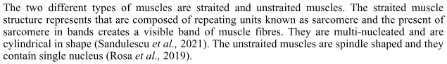
Sandulescu, T., Weniger, J., Philippou, S., Mücke, T., Naumova, E.A. and Arnold, W.H., 2021. Immunohistochemical evidence of striated muscle cells within midfacial superficial musculoaponeurotic system. Annals of Anatomy-Anatomischer Anzeiger, 234, p.151647.
Sefton, E.M. and Kardon, G., 2019. Connecting muscle development, birth defects, and evolution: An essential role for muscle connective tissue. Current topics in developmental biology, 132, pp.137-176.
Smith, D.A., 2018. The sliding-filament theory of muscle contraction. Springer International Publishing.
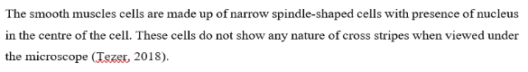
Tezer, M., 2018. Smooth muscle morphology in the nipple-areola complex. In Nipple-Areolar Complex Reconstruction (pp. 71-73). Springer, Cham.
Tieland, M., Trouwborst, I. and Clark, B.C., 2018. Skeletal muscle performance and ageing. Journal of cachexia, sarcopenia and muscle, 9(1), pp.3-19.
Watson, E.C. and Adams, R.H., 2018. Biology of bone: the vasculature of the skeletal system. Cold Spring Harbor perspectives in medicine, 8(7), p.a031559.
Wilke, H.J. and Volkheimer, D., 2018. Basic biomechanics of the lumbar spine. In Biomechanics of the Spine (pp. 51-67). Academic Press.
Bibliography
Li, E.W., McKee-Muir, O.C. and Gilbert, P.M., 2018. Cellular biomechanics in skeletal muscle regeneration. Current topics in developmental biology, 126, pp.125-176.
Liebsch, C. and Wilke, H.J., 2018. Basic biomechanics of the thoracic spine and rib cage. In Biomechanics of the Spine (pp. 35-50). Academic Press.
Tieland, M., Trouwborst, I. and Clark, B.C., 2018. Skeletal muscle performance and ageing. Journal of cachexia, sarcopenia and muscle, 9(1), pp.3-19.
Turgut, E. and Baltaci, G., 2020. Biomechanical principles of the exercise design. In Comparative Kinesiology of the Human Body (pp. 527-538). Academic Press.
Looking for further insights on Understanding Depression and Schizophrenia? Click here.


- 24/7 Customer Support
- 100% Customer Satisfaction
- No Privacy Violation
- Quick Services
- Subject Experts