Radioactive Decay and Emission of Alpha
- 10 Pages
- Published On: 14-12-2023
How the ranges of the penetrating power and the properties of alpha, beta, gamma and X-ray are related in their nature and properties:
Alpha, beta and gamma rays are discharged from the radioactive particle during substance radioactivity (Ménesguen and Lépy, 2021).
Alpha rays:
Alpha rays are made up of positively charged alpha particles. Each Alpha particle is the helium atom that consists of two neutrons and protons (Elregig, 2020). As compared to beta and gamma rays, Alpha particle has higher ionization capacity and minimum penetration power. Alpha rays have the high capability of ionizing the atoms that are situated close to them. Evidence suggests that due to the high ionizing capacity of Alpha rays the entry of Alpha particles cause serious damage insides the body.
Beta rays:
Beta rays consist of several beta particles that are extremely energetic electrons (Bakar et al. 2019). These electrons are released from the inner nucleus of a radioactive substance. During radioactivity, a neutron nucleus spits into three particles a proton, an electron and rapid emission of a beta particle. In the context of education dissertation help, it is important to understand how these particles interact with materials. The beta particle that is discharged from the protein nucleus is at a rapid pace. This is why beta particle has the higher penetration power as compared to the Alpha particles. On the other hand, as compared to the alpha particles the ionizing capacity of beta particles is lower
Gamma rays:

Gamma rays are the highest frequency electromagnetic spectrum that contains no mass. Gamma rays have the highest penetration power as compared to the alpha and bets rays. Gamma rays have the least ionizing ability (Faucher-Giguère, 2020). Despite the lower ionizing ability, it is difficult to resist the gamma rays entering the body.
X-ray:
The X-ray can also be called the X radiation which is the form of electromagnetic radiation. X-ray is a powerful wave of electromagnetic energy. The penetration power of X-ray increases with the increase of its energy and frequency (Somaily et al. 2020). On the other hand, the ionizing capacity of X-ray is lower than that of Alpha rays. As compared to beta and gamma rays the penetration power of X-ray is lower but higher than that of Alpha rays.
Safety procedures associated with using the alpha, beta, gamma and X rays:
Different safety procedure can be used to avoid hazards or accidents during using radioactive particles or electromagnetic waves such as alpha, beta, gamma and X rays. These safety procedures are as follows:
Reducing the exposure time:
The external hazards can easily be minimized by reducing the exposure to radioactive particles such as alpha, beta and gamma rays and the electromagnetic waves such as X-ray through proper planning, dry runs and well-constructed process of radionucleotide handing, it is possible to minimize the risk of explosion as well as the other associated hazards during using these radioactive an electromagnetic wave (Elregig, 2020).
Increasing distance:
By maintaining a standard distance between the operation or patients and radiation source it is possible to use electromagnetic rays safely (Zhao et al. 2020). The operator musty use the tools that have long handles thereby make a safe distance between the operation and the radioactive particles. Moreover, the operation must use the stands and clamps in terms of holding the radioactive substance safely.
Shielding:
By shielding the radiation sources, it is possible to reduce the intensity of the radiation from radioactive particles (Chaouch et al. 2018). In this entire process, the radiation is scatted by using the shield. Here the operator or the patient must stay always from the edge of the shield. In the case of using the gamma emitter, a shield of high thickness needs to use of because the gamma rays have high penetration capacity.
Use of barium meals in soft body imaging:
Barium meals
A barium meal is a diagnostic test that is used for examining the abnormalities in structure and function of the small duodenum, oesophagus and stomach by using the X-ray (Chaouch et al. 2018). Generally, an X-ray is able to highlight only the bones but not the soft tissues insides the inner inning of the internal body organs. For eliminating this problem, the clinical expert uses barium meal in which radiopaque salt, that medium barium sulphate is used to coat the inner lining of the target organs of the digestive tract which enables the expert to get the accurate X-ray imaging of the soft tissues of the target organs.
Operator:
Barium meal test is performed by radiologists which are expert in performing the electromagnetic imaging of the internal body tissues and organs.
Reason for this test:
A barium meal test is carried out to diagnose different disorders in the digestive tract. For example, hernia, constriction, masses or obstruction in the stomach and oesophagus are detected by using a barium meal test (Tanimori et al. 2017).
Risks;
The risk of this barium test is associated with how long the patients are exposed to the X-ray. The risk is similar to the risk associated with a patient’s exposure to any radioactive or ionizing agents (Yoshida et al. 2020). However, in the case of performing the barium meal test the time of patient's exposure to X-ray is minimum and the radiation is also low which contribute to very low risk for the patient. On the other hand, the barium liquid can easily be absorbed into the patient’s body thereby causing no harm to the patient.
Use of Gamma rays in imaging:
Gamma rays that are emoted from tiny radionucleotides are administered into the patient's body to get accurate medical diagnostic images (Tomono et al. 2017). The imaging using gamma rays is used in both court medicines and nuclear medicine. Gamma rays are used in different ranges of imaging to carry out effective medical diagnosis. Gamma rays are used in
Infection imaging
Tumour managing
Thyroid imaging
Brain imaging
Cardiac imaging
Diagnosis of the Alzheimer patient
Gamma rays are used in modern medical science in terms of producing both 2D and 3D images in the internal organs of the body. Scintigraphy is the process that is used while carrying out 2S gamma imaging in which the internal radionucleotides are used for the imaging
By using gamma rays 2D images are produced in nuclear medicine (Rangraz et al. 2020). Gamma rays are used in performing the full body bone scan. In this process, Gamma rays imaging assist the radiologist to examines the bone stricter, any animalistic in bone structural forms and the pathological aspects of the bone. In nuclear medicines, gamma rays imaging is used to examine, bone fracture, bone pain, infection in the bone, bone cancer and noncancerous lesion on bone.
Use of the internal process of the radiation in the treatment process
Internal radiation therapy is called brachytherapy which is associated with administering a higher dose of electromagnetic radiation on a particular target area as compared to the dose administered by the external rational therapy (Ning et al. C., 2019). The internal radiation source consists of a holder which is called the implant. The efficiency of this internal therapy does not depend on what types of impact have been used in the body. This process depends mainly on how perfectly the impact on places into the target organ inside or close to the tumour. The internal therapy poses the radiation on particulate body cells, therefore, causing fewer harms to the surrounding normal cells.
During performing the internal cavitary radiation the implant needs to be placed inside the uterus or the rectum thereby examining the inner tissues and cells inside the organs to determines any abnormalities in their functions (Kumar et al. 2021). During the intestinal radiation, the implant is placed near or inside the tumour rather than placing on the body cavity.
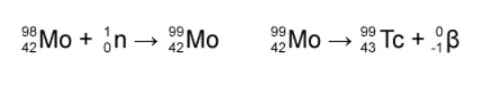
How technetium-99m is generated:
Technetium-99m is the metastable nuclear isotope of technetium-99 ((Famiglietti et al. 2017). Most commonly technetium-99m is also known as the reactive isotope which is used in tens of millions of clinical procedures. It emits gamma rays that have an energy of about 140 km V which is highly useful for detection as well as diagnosis of a pathological condition. Technetium-99m is generated by using neutrons with bombarding Molybdenum (98Mo). As a result, bombarding Molybdenum (98Mo) is generated. The produce is then pass through a bet decay process in which it passed its half-life of 66 hours thereby producing technetium (Sulieman et al. 2018). Ultimately Technetium-99m of generated which is now ready to be used in any medical procedure for the detection of internal tissues and organ. Similar to the other isotope of technetium, Technetium-99m is also unstable. Therefore Technetium-99m ends up producing Ruthenium-99 after undergoing the beta decay.
Use of iodine-131 in thyroid investigation:
Iodine-131 is the radioactive isotope that is widely used in modern medical science to treatment of thyroid cancer and production of thyroid imaging. As mentioned by Tanimori et al. (2017), the Environment Protection Agency, Iodine-131 has a very short half life span of only eight days. Therefore Iodine-131 undergoes complete decay within 30 days. This is why this isotope is used in many medical processes.
Hyperthyroidism:
Hyperthyroidism is the condition in which the thyroid gland is overactivated thereby secretion a high amount of thyroxin. As a result, there is a higher rate of metabolisms which leads to a faster reduction in body weight and an abnormal heart rate. In this condition, Iodine-131 is used to manage the condition. In the case of patients with hyperthyroidism, they are administered with Iodine-131 tablets which can easily be absorbed in the thyroid gland.
Thyroid Cancer:
Iodine-131 is widely used in treating thyroid cancer, in which this isotope is placed inside the cancerous cells of the thyroid gland by using the implant. The process this is used in this implantation is called brachytherapy (Yoshida et al. 2020). After implantation Iodine-131 emits gammas rays which are strongly imposed on only the cancerous cells but not on the surrounding normal cells. Iodine-131 destroy the cancerous cells of the thyroid gland
Thyroid imaging:
Iodine-131 acts as the radiotracer which is used to detect the abnormalities in the structure and functions of the different parts of the thyroid gland (Tomono et al. 2017). In this context, the Iodine-131 is administered into the patient's body through intramuscular pr intravenous injection. After entering into the body, the radiotracer emits the gamma-ray. Then the energy (gamma rays) is detected by using a gamma camera and the PET scanner. By using the computer, the absorbed amount of the radiotracer is measured and thus producing the images of the target parts of the thyroid gland.
Medical use of the parts of the electromagnetic spectrum:
Different electromagnetic rays are used in different clinical interventions such as detecting cancerous cells, determining the disorders in function and structure of internal organs and pathological characteristics of tissues and cells (Rangraz et al. 2020).
Gamma rays are used in producing the 2D and 3D imaging of different organs and soft tissues such as thyroid imaging, infection imaging, brain imaging and cardiac imaging (Famiglietti et al. 2017). Gamma rays are used in deleting the anomalistic in the function of the nerve cells in Alzheimer patients.
X rays are used is producing images of bones which assist radiologists to detect bines fractures, any malignancy in the bones and disorder in the bone function.
The electromagnetic spectrum is used not only for the detection of abnormal or cancerous cells but also in destroying the cells. In the case of treatment of thyroid cancer, Iodine-131 is used which after entering into the body completes its half-life and produce gamma rays that destroy the cancerous cells.

Use of the ultrasonic in medical imaging and treatment:
Ultrasonic radiation is used in treating joint fractures, joint pains and different types of tumours. This is used in detecting the types of tumours, whether they are benign or malignant (Tomono et al. 2017). Medica ultrasound is the widely used imaging techniques that are used in the clinical field to produces accurate images of bone, joints, blood muscles tendons and internal organs. Obstetric ultrasound is used in case of the pregnant women to detect the position of the fetus and proper functions and structure of internal glands such as the thyroid and pancreas
Dig deeper into Racism and Education with our selection of articles.
Reference list:
Bakar, N.F.A., Othman, S.A., Azman, N.F.A.N. and Jasrin, N.S., 2019, June. Effect of ionizing radiation towards human health: A review. In IOP Conference Series: Earth and Environmental Science (Vol. 268, No. 1, p. 012005). IOP Publishing.
Chaouch, M.A., Nacef, K., Ghannouchi, M., Khalifa, M.B., Chaouch, A., Abdelkafi, M., Jerbi, S. and Boudokhane, M., 2018. Choledochoduodenal fistula due to peptic duodenal ulcer diagnosed by X-barium meal study: interest of medical treatment. Pan African Medical Journal, 29(1), pp.1-4.
Elregig, R.A.E., 2020. Effect of X-rays and Gamma Rays on Tomato Seeds Growth. Sudan University of Science and Technology.
Famiglietti, R.M., Norboge, E.C., Boving, V., Langabeer, J.R., Buchholz, T.A. and Mikhail, O., 2017. Using discrete-event simulation to promote quality improvement and efficiency in a radiation oncology treatment center. Quality management in health care, 26(4), pp.184-189.
Faucher-Giguère, C.A., 2020. A cosmic UV/X-ray background model update. Monthly Notices of the Royal Astronomical Society, 493(2), pp.1614-1632.
Kumar, K.A., Balazy, K.E., Gutkin, P.M., Jacobson, C.E., Chen, J.J., Karl, J.J., von Eyben, R. and Horst, K.C., 2021. Association Between Patient Education Videos and Knowledge of Radiation Treatment. International Journal of Radiation Oncology* Biology* Physics, 109(5), pp.1165-1175.
Ménesguen, Y. and Lépy, M.C., 2021. COLEGRAM, a flexible user-friendly software for processing of ionizing radiation spectra. Nuclear Instruments and Methods in Physics Research Section A: Accelerators, Spectrometers, Detectors and Associated Equipment, 1003, p.165341.
Ning, M.S., Gomez, D.R., Shah, A.K., Kim, C.R., Palmer, M.B., Thaker, N.G., Grosshans, D.R., Liao, Z., Chapman, B.V., Brooks, E.D. and Tang, C., 2019. The insurance approval process for proton radiation therapy: a significant barrier to patient care. International Journal of Radiation Oncology* Biology* Physics, 104(4), pp.724-733.
Rangraz, E.J., Tang, X., Maleux, G., Dekervel, J., Van Cutsem, E., Verslype, C., Baete, K., Nuyts, J. and Deroose, C.M., 2020. Hybrid Imaging Reveals Improved Absorbed Dose from Selective Internal Radiation Treatment by Using an Anti-Reflux Catheter.
Sangro, B., Martínez‐Urbistondo, D., Bester, L., Bilbao, J.I., Coldwell, D.M., Flamen, P., Kennedy, A., Ricke, J. and Sharma, R.A., 2017. Prevention and treatment of complications of selective internal radiation therapy: expert guidance and systematic review. Hepatology, 66(3), pp.969-982.
Somaily, H.H., Algarni, H., Alraddadi, S., Rammah, Y.S., Nutaro, T. and Al-Buriahi, M.S., 2020. Mechanical, optical, and beta/gamma shielding properties of alkali tellurite glasses: role of ZnO. Ceramics International, 46(18), pp.28594-28602.
Sulieman, A., Elhag, B., Alkhorayef, M., Babikir, E., Theodorou, K., Kappas, C. and Bradley, D., 2018. Estimation of effective dose and radiation risk in pediatric barium studies procedures. Applied Radiation and Isotopes, 138, pp.40-44.
Tanimori, T., Mizumura, Y., Takada, A., Miyamoto, S., Takemura, T., Kishimoto, T., Komura, S., Kubo, H., Kurosawa, S., Matsuoka, Y. and Miuchi, K., 2017. Establishment of imaging spectroscopy of nuclear gamma-rays based on geometrical optics. Scientific Reports, 7(1), pp.1-12.
Tomono, D., Mizumoto, T., Takada, A., Komura, S., Matsuoka, Y., Mizumura, Y., Oda, M. and Tanimori, T., 2017. First on-site true gamma-ray imaging-spectroscopy of contamination near Fukushima plant. Scientific reports, 7(1), pp.1-10.
Yoshida, E., Tashima, H., Nagatsu, K., Tsuji, A.B., Kamada, K., Parodi, K. and Yamaya, T., 2020. Whole gamma imaging: a new concept of PET combined with Compton imaging. Physics in Medicine & Biology, 65(12), p.125013.
Zhao, P., Li, X., Tang, H., Ma, Y., Chen, B., Xing, W., Liu, K. and Yu, J., 2020. Improved high-temperature oxidation properties for Mn-containing beta-gamma TiAl with W addition. Oxidation of Metals, pp.1-16.


- 24/7 Customer Support
- 100% Customer Satisfaction
- No Privacy Violation
- Quick Services
- Subject Experts