Oblongata And Regulatory Mechanisms
Introduction
Heart rate refers to the number of complete contraction of the atria and ventricle per minute. Stroke volume refers to the average volume of blood pumped out of the left ventricle in a complete heartbeat (Kim et al.,2016). Cardiac output refers to the amount of blood pumped from the right or left ventricle per minute. The medulla oblongata is a special organ located in the hindbrain that uses both the nervous system and the endocrine system to control autonomic functions in the body such as breathing, heart activity (heart rate and stroke volume) and blood pressure functioning so as to maintain homeostasis (Antonio et al.,2014). Chemoreceptors inside the body fire action potential in stimulus changing chemical composition in the environment they are in, especially blood pH while baroreceptors detect pressure and stretch change in the area where they are located. These two resources influence the working of the medulla oblongata to maintain homeostasis while cycling. For those studying such complex physiological concepts and seeking healthcare dissertation help, understanding these mechanisms is crucial for developing comprehensive research. (Fig. 3).
Blood pressure regulation
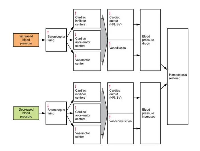
The nervous system detects blood pressure using baroreceptors. Vascular baroreceptors are located in sinuses at the swelling of the ascending aorta superior to the aortic valve (monitors pressure of blood going to the rest of the body and at the base of the internal carotid artery (monitor pressure of blood flowing to the head) and low pressure baroreceptors are on the walls of the vena cava and right atrium (Kim et al. 2016). This facilitates monitoring of blood pressure. When blood pressure rises above normal, 120/70 mmHg, baroreceptors increase action potential generated and fired to the medulla oblongata vasomotor center (Fig. 3). Parasympathetic stimulation of the heart increases reducing heart rate and cardiac output (CO) decreases. Furthermore, sympathetic stimulation of peripheral arterials decreases causing vasodilation leading to a decrease in blood pressure, decreasing cardiac preload and stroke volume (Richard et al., 2015). When blood pressure is below normal, baroreceptors decrease the generation and firing of action potential, increasing sympathetic stimulation which increases heart rate and CO is increased. Sympathetic stimulation of peripheral vessels is increased causing vasoconstriction which raises blood pressure and increases cardiac preload and stroke volume. (Fig. 1)

The medulla oblongata has three important regions, the cardio-accelerator center that is linked to sympathetic nerve, cardio-inhibitor center linked to the parasympathetic nerve and vasomotor center for the vascular system. Each of them is used depending on the activity the person is taking. The subject of this experiment was a 29-year-old female. She rested for the first six minutes of the experiment and her heart rate, blood pressure and oxygen percentage readings were taken. A normal adult has a heartbeat rate of 60-100 beats/minute (Kim et al. 2016). Her heartbeat reading was taken at 1-minute interval during the rest period and it was within the normal range with her lowest being 83 beats/minute and the highest at 92 beats/minute.
The normal blood pressure of an adult is 120/70 mmHg in systolic and diastolic pressure respectively and it is important to keep blood pressure within normal range (Ichinose et al.,2014). Resting blood pressure was taken twice. The first recording was 122/81 mmHg which is slightly above the normal systolic value. This was attributed to a possible increase in the sympathetic innervation. The second reading was 119/70 mmHg which was within normal range. The oxygen percentage concentration was fairly high all through the six minutes.
Cycling between minute 7 and 18 causes the heart rate to promptly increases. It rises until minute 9 where it reaches its highest of 170 beats/minute which is normal because in adults it rarely reaches 195 beats/minute compared to children which can reach 200 beats/minutes during exercise. The heart rate then fluctuates but remains fairly above 107 beats/minutes which is the lowest. Blood pressure also increases from 148/99 to 227/193 mmHg then to 210/165 mmHg. The oxygen percentage consumption remains relatively high despite its frequent fluctuations. At the onset of the cycling exercise there is an immediate increase in metabolic activity in the cell to produce more ATP energy for cycling. An increase in metabolic activity demands for more oxygen to be supplied and in the process more carbon (iv) oxide produced. An increase in its concentration lowers blood pH. Cycling is an isotonic exercise involving large muscle mass and in this case vasodilation occurs in the muscles involved for more oxygen supply and waste removal.
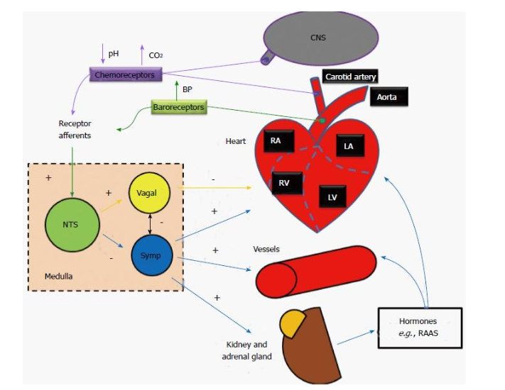
Blood pressure has to be regulated for enough oxygen to be supplied to remove carbon (iv) oxide and at the same time the perforating through the capillaries does not cause any damage. Vasodilation in the muscles due to vasodilators like adenosine and carbon (iv) oxide, reduces systemic vascular resistance (SVR) dropping the blood pressure which becomes a danger to the brain (Ichinose et al.,2014). Blood mechanically pumped by skeletal muscles to the heart and there is an increase in myocardial preload and stroke volume increases (Antonio et al.,2014). Once in the heart blood passes through the aorta and carotid arteries where chemoreceptors are located and the low pH fires a potential to the medulla oblongata. Mechanical pumping of the skeletal muscle pushes blood harder into the right atrium which is forced to stretch to accommodate the extra blood. An increase in myocardial preload distends the baroreceptors found in the aortic arch and carotid sinuses. Stretch receptors are part of the baroreceptors and they relay generated action potential to the rostral ventro-lateral medulla (RVLM). The medulla oblongata then processes the information sent from both the chemoreceptors and baroreceptors (Fig. 2).
Information is then relayed from the medulla oblongata through the accelerator nerve that uses the endocrine system. Noradrenaline is released from the nerve terminal and binds on the Beta 1 adrenergic receptor in the Sino Atrio-Node (SAN) conduction center (It dictates the rhythm of the heartbeat) to cause physiological changes such as increased intracellular calcium and also increases inotropic (force and speed of the heart), chronotropic (heart rate) and dromotropic (conduction speed) heart functions (Richard et al. 2015). This increases the heart rate and stroke volume hence cardiac output (CO) is increased countering the dropped SVR. The stroke volume needs to be increased for more CO hence blood reaches the exercising muscles to supply oxygen and glucose faster and take away carbon dioxide, lactate and heat from muscles faster. Stroke volume always increases with the intensity of the exercise.
The conduction of the atrio-ventricular node (AVN) conducting system increases. More blood is pumped to the lungs where carbon (iv) oxide diffuses out of blood into the lungs and more oxygen diffuses into blood and is transported to the skeletal muscles. The sympathetic nerve from the vasomotor center affects the tunica media of the arteries and veins. When the venous return is less than the CO, blood pressure in the venous blood vessels decreases. Baroreceptors in the thin part of vena cava and right atrium detect this and fire action potential to the medulla oblongata, increasing sympathetic stimulation which will cause vascular resistance, increasing pressure thus more venous return (Antonio et al.,2014). There is also an increased amount in cardiac preload resulting to a larger stroke volume. There is increased vascular resistance in abdominal viscera and non-active skeletal muscles so that more CO is directed to the active skeletal muscles where there is vasodilation.
During the recovery period the subject’s heart rate slowly began decreasing but fluctuated and rose a bit before decreasing once more but above the normal range, the lowest heart rate recorded within this time was 74 beats/min. Her blood pressure decreased from the previous to 143/93 mmHg before rising to 149/92 mmHg then reducing to 141/84 mmHg. The oxygen percentage consumption remained relatively high with varying values. After stopping the exercise, the medulla oblongata is gradually trying to reduce the sympathetic inputs and increases vagus nerve action (Antonio et al., 2014). The mechanical muscle pump has stopped which reduces the cardiac preload. The muscle cells however are still having a high metabolic rate which results to a low blood pH. The venous return is still vasoconstricted hence blood pressure is high despite the stopped mechanical pump (Coutsos et al.,2013). More carbon (iv) oxide diffuses out of blood and more oxygen is supplied to the muscle. Eventually, the chemoreceptors transmit the reduced acidity to the medulla oblongata which stimulates the vagus nerve that secretes acetylcholine to the SAN and through the M2 receptor cause a negative inotropic, chronotropic and dromotropic action, eventually reducing cardiac output. M3 receptors on the blood vessels also cause dilation of the constricted vessels, slowly reducing blood pressure (Coutsos et al.,2013).
In conclusion, the process of exercising induces parasympathetic withdrawal and causes sympathetic activation to enhance myocardial contractility and increasing heart rate and stroke volume increasing cardiac output. Parasympathetic nerves regulate basal metabolic functions. Chemoreceptors and baroreceptors are very crucial in the proper functioning of the medulla oblongata (Antonio et al.,2014).
Take a deeper dive into Obesity Trends in Childhoods with our additional resources.

REFERENCES
- Amann M., Blain G. M., Protor L. T., Sebranek J. J., Pegelow D. F., Dempsey J. A.. 2011. Implications of group III and IV muscle afferents for high intensity endurance exercise performance in humans. Journal of Physiology.
- Antonio C. L. Nobrega, Donal O’Leary, Bruno Moreira Silva, Elisabetta Marongui, Massimo F. Piepoli, Antonio Crisafulli. 2014. Neural Regulation of Cardiovascular Response to Exercise: Role of Central Command and Peripheral Afferents.
- Coutsos M., Sala-Mercado J. A., Ichinose M., Li Z., Dawe E. J., O’Leary D. S.. 2013. Muscle metaboreflex-induced coronary vasoconstriction limits ventricular contractility during dynamic exercise in heart failure. The American Journal of Physiology-Heart and Circulatory Physiology
- Dempsey J. A.. 2012. New perspectives concerning feedback influences on cardiorespiratory control during rhythmic exercise and on exercise performance. The Journal of physiology.
- Forster H. V., Haouzi P., Dempsey J. A.. 2012. Control of breathing during exercise. Comprehensive Physiology.
- Ichinose M., Maeda S., Kondo N., Nishiyasu T., 2014. Blood pressure regulation II: what happens when one system must serve two masters: oxygen delivery and pressure regulation? European Journal of Applied Physiology and Occupational Physiology.
- Kumar P., Prabhakar N. R. 2012. Peripheral chemoreceptors: function and plasticity of the carotid body. Comprehensive Physiology.
- Lombardi F., Stein P. K., Front Physiol, 2011. Origin of the heart rate variability and turbulence: an appraisal of autonomic modulation of cardiovascular function.
- Mann D. L., Zipes D. P., Libby P., Bonow R. O., 2014. Braunwald’s Heart Disease: Textbook of Cardiovascular Medicine. 10th edition. Philadelphia, Pennsylvania. Elsevier. Health Science Division.
- Marsac J., 2013. Heart rate variability: a cardiometabolic risk marker with public health indications. Bull Academy
- Metelka R., 2014. Heart rate variability- current diagnosis of the cardiac autonomic neuropathy. A review Biomed Pap Med Fac Univ Palacky Olomouc Czech Repub.
- Richard Gordan, Judith K., Lai Xie, 2015. World Journal of Cardiology: Autonomic and Endocrine control of cardiovascular function.
- Silva B. M., Fernandes I. A., Vianna L. C.. 2012. Welcome the carotid chemoreflex to the “neural control of the circulation during exercise” club. The Journal of Physiology.
- Stickland M. K., Morgan B. J., Dempsey J. A., 2011. Carotid chemoreceptor modulation of sympathetic vasoconstrictor outflow during exercise in healthy humans. Journal of Physiology.
- Thijssen D. H. J., Maiorana A. J., O’Driscoll G., Cable N. T., Hopman M.T.E, Green D. J. 2010. Impact of inactivity and exercise on the vasculature in humans. European Journal of Applied Physiology.
- Whyte J. J., Laughlin M. H.. 2010. The effect of acute and chronic exercise on the vasculature. Acta Physiologica .


- 24/7 Customer Support
- 100% Customer Satisfaction
- No Privacy Violation
- Quick Services
- Subject Experts