Understanding Cell Biology Basics
Introduction
The cell biology informs the basic functional, structural and biological unit of life present in all living organisms which is considered as the building block of the life. In this assignment, a detailed overview of the cell biology of different cells and its functioning is to be discussed. For this purpose, initially the basic cell structure and its organelles are explained. Thereafter, the way metabolism in the cell occurs through different methods and techniques are discussed. Further, the ways cells function to dive and grow through different process are explained.
Section 1
Discuss the selected characteristics of living cells.
Cellular Reproduction: The characteristics of living cell includes that they are able to reproduce though cell division through the process of binary fission where two identical daughter cells are formed out of a single parent cell, mitosis where chromosome present in the nucleus of the cell is used and they divide to form genetical identical copies of daughter cells and meiosis that occurs in the living sex cells where number of chromosome in the cell is halved (Phillips et al., 2012).
Support Cell metabolism: The living cells use Adenosine Triphosphate (ATP) as the energy source for driving metabolism in the body. The living cells are able to develop effective adaption according to the environmental condition and are self-sufficient in performing actions (Sahmani and Aghdam, 2017).
Homeostasis: The living cells support homeostasis which is the process of creating a stable environment within the body such as constant temperature, normal blood sugar level, normal breathing rate and others. The living cells show effective response to stimuli which is simple reaction to any external or internal force towards the cells (Zhao et al. 2020).
Cellular Organisation: The functional characteristics of living cells include presence of well-developed cellular organization. This means presence of all the organelles such as mitochondria, nucleus, lysosomes, endoplasmic reticulum and others in active state (Tang et al., 2020).
Cell response to stimuli: The living cells are seen to effectively respond to any internal or external stimuli by changing their shape, making movement, altering cell integrity and others (Tang et al., 2020).
Compare and contrast prokaryotic and eukaryotic cells, and explain the impact that viruses have on them.


The virus inhabiting the mammalian host is subdivided into eukaryotic viruses that infect the other eukaryotic cells and host cells and bacteriophage that infects the prokaryotic cells. The eukaryotic virus mainly coming in contact with the host cells acts to insert own genetic material in the cell and negatively affect the tissue-specific immunity which results individuals to develop hindered health condition and fever with complication in body functioning out of reduced immunity (Aiewsakun and Simmonds, 2018). The bacteriophage mainly affects the prokaryotic cell in which they enter to cause reproduction and finally break out from them to release new viruses in turn killing the bacteria (Zhang et al., 2017).
Discuss eukaryotic sub-cellular structure and organelles.
The eukaryotic sub-cellular structure and organelles are as follows:
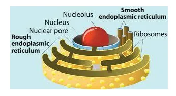
Nucleus: It is the largest organelle that has been surrounded by double-fold of nuclear membrane which has presence of pores so that it can control the entry and exit of ribosomes and RNA in and out of the nucleus. The nucleus has the function to coordinate cell division and synthesis as it contains the genetic material. The nucleoli are smaller bodies present in the nucleus and the gel-like matrix in the nucleus is known as nucleoplasm in which all the nuclear components and genetic material are submerged (Lykov et al., 2017).

Cytoplasm: The cytoplasm is the thick solution present inside the cell and fills in which all the internal cellular organelles are present. It mainly contains salts, water and proteins in its composition. The cytoplasm mainly acts as buffer in protecting the cell’s genetic material and provides environment for the cellular organelles to avoid getting damaged during collision or movement (Gabaldón Estevan and Pittis, 2016)

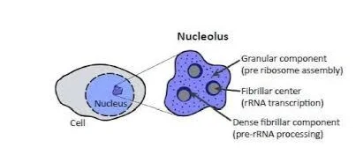
Nucleolus: It is present inside the nucleus and plays the role of supporting manufacture of ribosome in the nucleus of the cell (Tjondro et al., 2019)

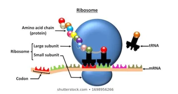
Ribosome: These are small organelles which are present in numerous amount within the cell and acts as key sites of protein synthesis. They function to decode message in forming peptide bonds and ribosome has two different subunits in which one is larger and another smaller. Each of these subunits are made of ribosomal proteins (Tjondro et al., 2019)

Endoplasmic Reticulum (ER): The endoplasmic reticulum is of two types that are smooth ER and rough ER. The smooth ER mainly plays the role of supporting lipid synthesis and manages transport of the produced lipid whereas the rough ER shows responsibility in processing proteins developed by ribosomes along with modifying them for adding carbohydrate to it (Lykov et al., 2017).

Golgi body: It is mainly stack of small flat sacs that receive protein from the ER and acts to modify them to be repacked to be used for cellular secretion. They act in processing and packing proteins for exporting them to the cell (Lykov et al., 2017).

Lysosome: The lysosomes are membrane-bound cellular organelles which contains digestive enzymes inside them. It acts to cause waste disposal with the help of vacuoles. They act in destroying the bacteria and viruses that enters the cell for protecting it from damage (Lykov et al., 2017)


Section 2
The role of the cell membrane in regulating how nutrients are gained and waste products lost.
The cell membrane is mainly present surrounding the cytoplasm on the living cell which allows it to physically separate the intracellular organelles of the cell from external environment. The role played by them in regulating gain of nutrients and release of waste materials are as follows:
Passive osmosis and diffusion: The small molecules and ions like oxygen and carbon dioxide are able to move in and out of the plasma membrane with the help of diffusion that is mainly a passive action. Since the plasma membrane acts as barrier to certain molecules, the diffusion occurs across two sides of the membrane. In the passive diffusion, the plasma membrane allows the small ions and molecules to pass freely from low concentration to higher concentration to create equilibrium. The passive osmosis used by plasma membrane in gaining nutrients includes passage of molecules from low concentration to higher concentration across a semi-permeable membrane (Qi et al., 2018)
Trans-membrane protein channels: These channels extend from the membrane through the lipid bilayer and they functions to support gain of nutrients and transport across both sides of the membrane. The nutrients like amino acids and sugar molecules diffuse passively with the help of aquaporins in facilitated diffusion or are pumped by trans-membrane transporters across the membrane. The protein channels allow only selected substances to be passed through the cell (Alcaraz and Queralt-Martín, 2016).
Endocytosis: The endocytosis is the approach of absorbing molecules by the cells by engulfing them. In this process, the plasma membrane initially develops a small deformation inside the cell membrane through the process known as invagination to allow the substances to be transported in the invasion for engulfing them. The proteins present in the outer part of the membrane acts as receptor and support clustering to depress the membrane to eventually promote accumulation of lipids and proteins in the inner side of the membrane. The deformation caused pinches the membrane off and inside the cell to create a vesicle which contains the substance to be captured. This requires energy and therefore it is regarded as active transport (Li et al., 2016).
Exocytosis: The exocytosis is discarding materials outside the cell and in this process the vesicle formed inside the membrane fuses with the plasma membrane to extrude the materials out from the cell. It mainly occurs to move waste materials outside the cells. The process mainly initiate with the Golgi-body forming vacuoles present with undigested and waste materials inside the cell membrane that are later transported to the edge of the membrane for fusion to led the materials out of the cell (Mohammadi et al., 2018, Dix et al., 2018).
How animal cells use nutrients to provide the energy for growth, movement, and cell division.
The living animal cells require source of energy for performing growth, cell division movement and others which is provided to them from the storage of chemical energy in the form of ATP that is made up in the process of cellular respiration (Zhang et al., 2020). Cellular respiration is the process through which the living cell convert glucose in the form of molecules and release energy. The cellular respiration that leads to the production of ATP occurs in two stages that are glycolysis and Krebs cycle (Kornberg et al., 2018). The glycolysis occurs within the cytosol present inside the cytoplasm and in this process mainly splitting of glucose molecule occurs in different steps. In the first stage, the D-glucose breaks into glucose-6-phosphate with the action of enzyme hexokinase. The second step is change of glucose6-phosphate into fructose-6-phosphate with the action of glucose phosphate isomerase. The fructose-6-phosphate is changed into fructose-1,6-phosphate with magnesium as cofactor and phosfructokinase as enzyme. The fructose-1,6-phosphate is then broken down into dihydroxyacetone phosphate (DHAP) and glyceraldehyde 3-phosphate (GAP) through the action of enzyme aldolase. This follows inter-conversion of GAP and DHAP with action of enzyme triosephosphate isomerase. The dehydrogenation of GAPDH occurs and inorganic phosphate is added to glyceraldehyde 3-phosphate to produce 1,3-bisphosphoglycerate. In the following step, a phosphate group from the 1,3-bisphosphoglycerate is tranfered to ADP in forming ATP and 3-phosphoglycerate. Further, 2-phosphoglycerate is formed where with the action of enolase phosphoenolpyruvic acid (PEP) is produced. The pyruvate kinase acts of PEP to displace a phosphate to form ATP and pyruvic acid that are end products of glycolysis (Park et al., 2020). The kerbs cycle initiates with condensation of the acetyly-coA with oxaloacetate in forming citrate and the process uses citrate synthase as the enzyme. The acetyly-coA is mainly produced from the pyruvate developed in the glycolysis which on entering the mitochondria get transformed into acetyl-coA as resulting compound with release of one molecule of CO2 that is later picked by coenzyme A (coA). The citrate is rearranged to isocitrate with the action of acontinase as enzyme. In the following process, the isocitrate is changed to α-ketoglutarate with action of isocitrate dehydrogenase as catalyst. The α-ketoglutarate form succinyl-coA with removal of CO2 molecule and addition of CoA that is later removed in forming succinate from succinyl-coA. The succinate is further oxidized to form fumarate which later form L-malate in reversible reaction where the malate produce oxaloacetate initiating citric acid cycle by malate dehydrogenase (Liu et al., 2019).


The role of nucleic acids in the nucleus and cytoplasm.
There are mainly two types of nucleic acid which are:
Ribonucleic acid (RNA)
Deoxyribonucleic acid (DNA) (Choi et al., 2018)
The Ribonucleic acid (RNA) (Choi et al., 2018)
The Ribonucleic acid (RNA) is single-stranded nucleotide polymer which has three forms that are Ribosomal RNA (rRNA), Messenger RNA (mRNA) and Transfer RNA (t-RNA). The rRNA play the role of directly catalyzing steps in synthesis of protein so that amino acids are bound together to develop a protein molecule. The rRNA is also seen to bind with tRNA as well as other accessory molecules for supporting protein synthesis (Choi et al., 2018). The mRNA act to play the role of carrying the blueprint of the DNA of the cell to the ribosomes that are the machines which synthesize proteins. Thus, they act as intermediatory between the NA and protein to direct synthesis (Zhang and Jia, 2018). The tRNA has the role of decoding the message present in the mRNA sequence for protein synthesis. In this process, they function to chemically link with the particular amino acid alog with correct bae-pair of the mRNA through a coon o that the specific amino acid is able to be specifically attached growing peptide chain. During translation, the tRNA functions at specific sites within the ribosome in the process of synthesing a protein from mRNA molecule (Peschek and Walter, 2019). The DNA is the nucleotide which stores specific instruction for making other large molecules that are proteins. The four key roles of DNA are:
Replication: The DNA is double-stranded nucleotide where each of the base in one strand bind with complementary base present on the other complementary strand. During cell division, the DNA strands are to be replicated to make original copies so that the daughter cells receive the information (Morocz et al., 2017).
Encode information: The DNA contains four different bases that are adenine, guanine, cytosine and thymine. The adjacent trio of the bases in the DNA acts to provide place for specific amino acid in the proteins (Chen et al., 2016).
Mutation and Recombination: The DNA has the role to create evolution in species over time which they execute by addition or alteration of bases in the structure to form mutation. They also play the role to combine existing bases in the strands to form new DNA strand (Chen et al., 2016).
Gene expression: The DNA plays the role of supporting gene expression as they have genetic code for specific molecules to be produced present in them (Chen et al., 2016).
Discuss the synthesis of proteins
The protein synthesis mainly occurs in the ribosomes of the cells. There are two stages of protein synthesis which are translation and transcription. The transcription is the initial step of protein synthesis which the genetic information from the DNA is transferred to the mRNA. The steps in transcription are as follows:


The process of translation initiates after the transcription which has the following stages:
Initiation: In this stage, the mRNA which enters the ribosome is read by it to direct the tRNA bring correct sequence of amino acids for the mRNA in forming new strand of RNA. The ribosomes that are mainly out of rRNA has two subunits one small and another large. The reading initiate in the 5’-3’ manner for the mRNA where initially a small subunit is bond at the 5’ end on reaching the start codon that is AUG. The larger subunit and the initiator tRNA carrying methionine binds to the P site of the ribosome (Kearse and Wilusz, 2017).
The second stage is elongation in which one codon is shifted at a time leading to catalyze each process that are occurring on specific three sites at the mRNA. In each of the steps, tRNA enters and departs by adding amino acid according to the present sequence in the mRNA. The energy required for the bonding is derived from GTP that is similar to ATP. In brief, the ribosomes are also found to develop interaction with other RNA molecules in making polypetide chains because of the bonding of peptide formed in between amino acids. The three sites of ribosome mainly A, P and E show participation in the translation process (Chu et al., 2016).
The termination of the elongation occurs with encountering stop codon (UAA or UGA or UAG). On meeting the stop codon, the growing polypeptide is released with the assistance of releasing factors and the subunits of the ribosome dissociates leaving the mRNA (Chu et al., 2016).

Section 3
The generation of specialised tissues from embryonic stem cells.
The generation of specific tissues occurs from the embryonic stem cell as it is an undifferentiated cell with no specific indication for forming specific tissue. The embryonic stem cells are mainly obtained from the embryo at the initial stage of development before the implantation of the embryo occurs in the uterus. The fertilization mainly occurs within the oviduct and in some time series of cleavage division occurs with the progress of the embryo travelling from the oviduct to the uterus. The blastomeres at the cleavage-state embryo are found to be undifferentiated and show no confirmed action of any differentiated cell. The initial differentiation of the blastomeres occurs at the five day of development in human in which an outer layer of cell that shows commitment to form placenta separates from the inner mass of the cell (Sozen et al., 2018). The inner cell mass (ICM) has the potential to produce any nature of body but with implantation their power gets depleted because they are to differentiate into other type of cells with limited development potential. In contrast, if ICM can be removed from the normal embryonic environment and are cultured in well-maintained condition they show the ability to proliferate as well as replicate identity with the power to develop into any form or type of cell in the human body (Mattei et al., 2019). In the current condition, stem cell therapy is used in which by generating new cells, they are transplanted in the body to replace damaged cells. There are certain signals in the body which direct the nature of specialized cells to be prepared by the embryonic stem cell. The scientist by introducing such signals can make the stem cell show preparation to turn on genes for formation of specialized tissues (Kelava and Lancaster, 2016).

The process of interphase and factors that initiate cell division, and their importance
The interphase is the resting stage of the cell division and in this phase continuous synthesis of RNA and protein occurs allowing the cell to be healthy and grow in size to be fit for entering cell division. The importance of interphase is mainly to prepare the cell to enter cell division (Pecinka et al., 2020). The stages of interphase are:

The factors initiating cell division:
The release of growth-promoting hormones like HGH is able to influence initiation of cell division whereas its lack of presence in adequate amount would result in inhibiting cell division (Pecinka et al., 2020).
The presence of G cyclins allows cell growth and coordinate new cell to enter the cell cycle. The binding of the S-cyclins to Cdk induces DNA replication making cells to enter cell division (Pecinka et al., 2020).
How the same genetic information is received by each daughter cell.
The same genetic information is received by the daughter cells in the process of mitosis. The step of mitosis through which gradually the same genetic material is divided to be provided to each daughter cells includes:
Prophase: During the initiation of the cell division, the genetic material present in the cell is loosely packed in the form of chromatin. For the progress of the cell to divide, the chromatin material is required to be tightly packed for which it initiate to get condensed to form chromosome. Moreover, formation of mitotic spindle is seen that are structures developed from the centrosome. In mitosis, there are two centrosomes which initiate to move to opposite ends in the prophase (Keyormarsi et al., 2018).
Prometaphase: In this phase, the chromosome is found to be entirely condensed to form chromosome and the nuclear envelop initiate to get dissolved for allowing spindle fibers to be attached to the chromosomes. The mitotic spindles formed are attached at the kinetochore which is the area near the centromere (Keyormarsi et al., 2018).
Metaphase: The chromosomes are found to get aligned in a single line in this stage. Moreover, the centrosomes are found to be aligned near the metaphase plate with the kinetochores attached to them. It is required to ensure that the sister chromatids split effectively between two daughter cells without any error (Keyormarsi et al., 2018).

Anaphase: In this phase, the sister chromatids are to be pulled exactly in the opposite sides and the fibers are found to contract so that the chromatids are broken at the centromere to be moved two opposite plates formed in the cell. The spindle fibers are seen to elongate to prepare the cell for dividing (Keyormarsi et al., 2018).
Telophase: In this phase, the cells are found to have elongated and nearly finished to divide. The cells features present at the initiation of the division are found to reappear such as nuclear membrane and others. The chromosomes are found to become condensed and spindle fibers are broken creating two separate daughter ells (Keyormarsi et al., 2018).

Compare and contrast cancer cells with normal cells
The different between cancer and normal cells are as follows:


Conclusion
The above discussion mentions that living cell are able grow and divide, perform metabolism, support energy production and others. The key organelles are nucleus, mitochondria, cytoplasm and others. The metabolism in cell occurs through various processes such as glycolysis and Krebs cycle along with the transport of nutrients in and out of the cell is supported by active transport, diffusion and others. The cells divide through the process of mitosis and meiosis.
Recommendations
According to the discussed information, it is recommended that to ensure living cell performs effective metabolism proper glycolysis and Krebs cycle mechanism is to be supported. It is recommended that to identify living cells, the organelles are to be thoroughly analysed under the microscope to determine its life. It is also recommended that to differentiate between normal and cancerous cells, initially the nature of growth efficiency of the cells is to be considered an uncontrolled growth would indicate cancer cells whereas a controlled growth indicates normal cells.
References
- Aiewsakun, P. and Simmonds, P., 2018. The genomic underpinnings of eukaryotic virus taxonomy: creating a sequence-based framework for family-level virus classification. Microbiome, 6(1), p.38.
- Alcaraz, A. and Queralt-Martín, M., 2016. On the different sources of cooperativity in pH titrating sites of a membrane protein channel. The European Physical Journal E, 39(3), pp.1-6.
- Chen, S. and Zhao, Y., 2018. Circular RNAs: Characteristics, function, and role in human cancer. Histology and histopathology, 33(9), p.887.
- Chen, B., Le, W., Wang, Y., Li, Z., Wang, D., Ren, L., Lin, L., Cui, S., Hu, J.J., Hu, Y. and Yang, P., 2016. Targeting negative surface charges of cancer cells by multifunctional nanoprobes. Theranostics, 6(11), p.1887.
- Choi, J., Indrisiunaite, G., DeMirci, H., Ieong, K.W., Wang, J., Petrov, A., Prabhakar, A., Rechavi, G., Dominissini, D., He, C. and Ehrenberg, M., 2018. 2′-O-methylation in mRNA disrupts tRNA decoding during translation elongation. Nature structural & molecular biology, 25(3), pp.208-216.
- Danisovic, L., Oravcova, L., Krajciova, L., Varchulova Novakova, Z., Bohac, M., Varga, I. and Vojtassak, J., 2017. Effect of long-term culture on the biological and morphological characteristics of human adipose tissue-derived stem cells. J Physiol Pharmacol, 68(1), pp.149-158.
- Dix, C.L., Matthews, H.K., Uroz, M., McLaren, S., Wolf, L., Heatley, N., Win, Z., Almada, P., Henriques, R., Boutros, M. and Trepat, X., 2018. The role of mitotic cell-substrate adhesion re-modeling in animal cell division. Developmental cell, 45(1), pp.132-145.
- Kearse, M.G. and Wilusz, J.E., 2017. Non-AUG translation: a new start for protein synthesis in eukaryotes. Genes & development, 31(17), pp.1717-1731.
- Little, A.C., Sham, D., Hristova, M., Danyal, K., Heppner, D.E., Bauer, R.A., Sipsey, L.M., Habibovic, A. and Van Der Vliet, A., 2016. DUOX1 silencing in lung cancer promotes EMT, cancer stem cell characteristics and invasive properties. Oncogenesis, 5(10), pp.e261-e261.
- Lykov, K., Nematbakhsh, Y., Shang, M., Lim, C.T. and Pivkin, I.V., 2017. Probing eukaryotic cell mechanics via mesoscopic simulations. PLOS Computational Biology, 13(9), p.e1005726.
- Mohammadi, A.S., Li, X. and Ewing, A.G., 2018. Mass spectrometry imaging suggests that cisplatin affects exocytotic release by alteration of cell membrane lipids. Analytical chemistry, 90(14), pp.8509-8516.
- Pecinka, A., Chevalier, C., Colas, I., Kalantidis, K., Varotto, S., Krugman, T., Michailidis, C., Vallés, M.P., Muñoz, A. and Pradillo, M., 2020. Chromatin dynamics during interphase and cell division: similarities and differences between model and crop plants. Journal of experimental botany, 71(17), pp.5205-5222.
- Phillips, R., Kondev, J., Theriot, J. and Garcia, H., 2012. Physical biology of the cell. Garland Science.
- Qi, L., Hu, Y., Liu, Z., An, X. and Bar-Zeev, E., 2018. Improved anti-biofouling performance of thin-film composite forward-osmosis membranes containing passive and active moieties. Environmental science & technology, 52(17), pp.9684-9693.
- Simmonds, P., Becher, P., Bukh, J., Gould, E.A., Meyers, G., Monath, T., Muerhoff, S., Pletnev, A., Rico-Hesse, R., Smith, D.B. and Stapleton, J.T., 2017. ICTV virus taxonomy profile: Flaviviridae. The Journal of general virology, 98(1), p.2.
- Tjondro, H.C., Loke, I., Chatterjee, S. and Thaysen‐Andersen, M., 2019. Human protein paucimannosylation: cues from the eukaryotic kingdoms. Biological Reviews, 94(6), pp.2068-2100.
- Zhang, J., Gao, Q., Zhang, Q., Wang, T., Yue, H., Wu, L., Shi, J., Qin, Z., Zhou, J., Zuo, J. and Yang, Y., 2017. Bacteriophage–prokaryote dynamics and interaction within anaerobic digestion processes across time and space. Microbiome, 5(1), pp.1-10.
- Zhao, B., Katuwawala, A., Uversky, V.N. and Kurgan, L., 2020. IDPology of the living cell: intrinsic disorder in the subcellular compartments of the human cell. Cellular and Molecular Life Sciences, pp.1-15.
- Herce, H.D., Schumacher, D., Schneider, A.F., Ludwig, A.K., Mann, F.A., Fillies, M., Kasper, M.A., Reinke, S., Krause, E., Leonhardt, H. and Cardoso, M.C., 2017. Cell-permeable nanobodies for targeted immunolabelling and antigen manipulation in living cells. Nature chemistry, 9(8), p.762.
- Van Niel, G., d'Angelo, G. and Raposo, G., 2018. Shedding light on the cell biology of extracellular vesicles. Nature reviews Molecular cell biology, 19(4), p.213.
- 24/7 Customer Support
- 100% Customer Satisfaction
- No Privacy Violation
- Quick Services
- Subject Experts


